Efecto de la temperatura sobre la porosidad de andamios de hidroxiapatita y su uso en ingeniería de tejidos
DOI:
https://doi.org/10.37636/recit.v34213221Palabras clave:
Hidroxiapatita, andamios porosos, método de lixiviación de salesResumen
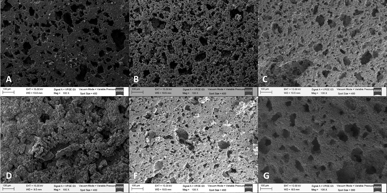
La búsqueda de un reemplazo óseo adecuado es de gran importancia debido a la dificultad de utilizar trasplantes autólogos. Es por esto, que el objetivo de este trabajo es comparar el efecto de la temperatura sobre la porosidad y el diámetro promedio de poro fabricados con el método de lixiviación de sales, siendo sinterizados desde ~150 a 1000 °C. Los andamios fabricados de hidroxiapatita fueron evaluados con difracción de rayos X (XRD). El potencial zeta fue evaluado a diferentes temperaturas. Los especímenes fueron caracterizados utilizando microscopia electrónica de barrido (SEM) y análisis Raman. Los resultados mostraron que la porosidad importante (57%) y tamaño de poro (49 µm) ocurren con un tratamiento térmico superior a ~ 850 °C para andamios que fueron pre-sinterizados a 1050 °C.
Descargas
Citas
K. Yang et al., “β-Tricalcium phosphate/poly (glycerol sebacate) scaffolds with robust mechanical property for bone tissue engineering,” Mater. Sci. Eng. C, vol. 56, pp. 37–47, Nov. 2015. http://dx.doi.org/10.1016/j.msec.2015.05.083. DOI: https://doi.org/10.1016/j.msec.2015.05.083
Z. Sheikh, C. Sima, and M. Glogauer, “Bone Replacement Materials and Techniques Used for Achieving Vertical Alveolar Bone Augmentation,” Materials, vol. 8, no. 6. 2015. http://dx.doi.org/10.3390/ma8062953. DOI: https://doi.org/10.3390/ma8062953
J. Lindhe. Periodontologia Clinica E Implantologia Odontologica. Médica Panamericana; 2009. https://books.google.com.mx/books?id=69zuJ1qspGwC.
L. J. Villarreal-Gómez et al., “Biocompatibility evaluation of electrospun scaffolds of poly (L-Lactide) with pure and grafted hydroxyapatite,” J. Mex. Chem. Soc., vol. 58, no. 4, pp. 435–443, 2014. http://dx.doi.org/10.29356/jmcs.v58i4.53. DOI: https://doi.org/10.29356/jmcs.v58i4.53
G. Hannink and J. J. C. Arts, “Bioresorbability, porosity and mechanical strength of bone substitutes: What is optimal for bone regeneration?” Injury, vol. 42, pp. S22–S25, Dec. 2016. http://dx.doi.org/10.1016/j.injury.2011.06.008. DOI: https://doi.org/10.1016/j.injury.2011.06.008
C. T. I. Reviews, Biomaterials Science, An Introduction to Materials in Medicine. Cram101, 2016. https://books.google.com.mx/books?id=TdrIAQAAQBAJ.
R. Bosco, J. Van Den Beucken, S. Leeuwenburgh, and J. Jansen, “Surface Engineering for Bone Implants: A Trend from Passive to Active Surfaces,” Coatings, vol. 2, no. 3. 2012. http://dx.doi.org/10.3390/coatings2030095. DOI: https://doi.org/10.3390/coatings2030095
L. J. Villarreal-Gómez et al., In vivo biocompatibility of dental scaffolds for tissue regeneration, vol. 976. 2014. http://dx.doi.org/10.4028/www.scientific.net/AMR.976.191. DOI: https://doi.org/10.4028/www.scientific.net/AMR.976.191
K. Kuroda and M. Okido, “Hydroxyapatite Coating of Titanium Implants Using Hydroprocessing and Evaluation of Their Osteoconductivity,” Bioinorg. Chem. Appl., vol. 2012, no. 1, pp. 1–7, 2012, doi: http://dx.doi.org/10.1155/2012/730693. DOI: https://doi.org/10.1155/2012/730693
C. Berger et al., “Ultrathin Epitaxial Graphite: 2D Electron Gas Properties and a Route toward Graphene-based Nanoelectronics,” J. Phys. Chem. B, vol. 108, no. 52, pp. 19912–19916, Dec. 2004. http://dx.doi.org/10.1021/jp040650f. DOI: https://doi.org/10.1021/jp040650f
L. Chen, Y. Tang, K. Wang, C. Liu, and S. Luo, “Direct electrodeposition of reduced graphene oxide on glassy carbon electrode and its electrochemical application,” Electrochem. commun., vol. 13, no. 2, pp. 133–137, Feb. 2011. http://dx.doi.org/10.1016/j.elecom.2010.11.033. DOI: https://doi.org/10.1016/j.elecom.2010.11.033
J. Shi, X. Li, G. He, L. Zhang, and M. Li, “Electrodeposition of high-capacitance 3D CoS/graphene nanosheets on nickel foam for high-performance aqueous asymmetric supercapacitors,” J. Mater. Chem. A, vol. 3, no. 41, pp. 20619–20626, 2015. http://dx.doi.org/10.1039/C5TA04464B. DOI: https://doi.org/10.1039/C5TA04464B
A. Di Luca et al., “Gradients in pore size enhance the osteogenic differentiation of human mesenchymal stromal cells in three-dimensional scaffolds,” Sci. Rep., vol. 6, p. 22898, Mar. 2016. http://dx.doi.org/10.1038/srep22898. DOI: https://doi.org/10.1038/srep22898
C. M. Murphy and F. J. O’Brien, “Understanding the effect of mean pore size on cell activity in collagen-glycosaminoglycan scaffolds,” Cell Adh. Migr., vol. 4, no. 3, pp. 377–381, Dec. 2010. http://dx.doi.org/10.4161/cam.4.3.11747. DOI: https://doi.org/10.4161/cam.4.3.11747
P. Kasten, I. Beyen, P. Niemeyer, R. Luginbühl, M. Bohner, and W. Richter, “Porosity and pore size of β-tricalcium phosphate scaffold can influence protein production and osteogenic differentiation of human mesenchymal stem cells: An in vitro and in vivo study,” Acta Biomater., vol. 4, no. 6, pp. 1904–1915, Nov. 2008. http://dx.doi.org/10.1016/j.actbio.2008.05.017. DOI: https://doi.org/10.1016/j.actbio.2008.05.017
L. D. Guillen-Romero et al., “Synthetic hydroxyapatite and its use in bioactive coatings,” J. Appl. Biomater. Funct. Mater., vol. 17, no. 1, 2019. http://dx.doi.org/10.1177/2280800018817463. DOI: https://doi.org/10.1177/2280800018817463
J. Wosek, “Fabrication of Composite Polyurethane/Hydroxyapatite Scaffolds Using Solvent-Casting Salt Leaching Technique,” Advances in Materials Science, vol. 15. p. 14, 2015. http://dx.doi.org/10.1515/adms-2015-0003. DOI: https://doi.org/10.1515/adms-2015-0003
S. Fragoso Angeles, R. Vera-Graziano, G. L. Pérez González, A. L. Iglesias, L. E. Gómez Pineda, and L. J. Villarreal-Gómez, “Síntesis y Caracterización de Hidroxiapatita Sintética para la Preparación de Filmes de PLGA/HAp con Potencial Uso en Aplicaciones Biomédicas,” ReCIBE, vol. 7, no. 2, pp. 93–116, 2018. http://recibe.cucei.udg.mx/ojs/index.php/ReCIBE/article/view/94. DOI: https://doi.org/10.32870/recibe.v7i2.94
S. Koutsopoulos, “Synthesis and characterization of hydroxyapatite crystals: a review study on the analytical methods”. J Biomed Mater Res. Vol. 62, Pp. 600–612, 2002. https://doi.org/10.1002/jbm.10280 DOI: https://doi.org/10.1002/jbm.10280
A.F. Khan, M. Awais, A.S. Khan, “Raman spectroscopy of natural bone and synthetic apatites”. Appl Spectrosc Rev. Vol. 48, Pp. 329–355, 2013. https://doi.org/10.1080/05704928.2012.721107 DOI: https://doi.org/10.1080/05704928.2012.721107
G.R. Sauer, W.B. Zunic, J. R. Durig, “Fourier transform Raman spectroscopy of synthetic and biological calcium phosphates”. Calcif Tissue Int. Vol. 54, Pp. 414, 1994. https://doi.org/10.1007/BF00305529 DOI: https://doi.org/10.1007/BF00305529
A.-J. Wang et al., “Effect of sintering on porosity, phase, and surface morphology of spray dried hydroxyapatite microspheres,” J. Biomed. Mater. Res. Part A, vol. 87A, no. 2, pp. 557–562, 2008, http://dx.doi.org/10.1002/jbm.a.31895. DOI: https://doi.org/10.1002/jbm.a.31895
S. V Dorozhkin, “Calcium orthophosphate deposits: Preparation, properties and biomedical applications,” Mater. Sci. Eng. C, vol. 55, pp. 272–326, Oct. 2015, doi: http://dx.doi.org/10.1016/j.msec.2015.05.033. DOI: https://doi.org/10.1016/j.msec.2015.05.033
C. F. C. Brown, J. Yan, T. T. Y. Han, D. M. Marecak, B. G. Amsden, and L. E. Flynn, “Effect of decellularized adipose tissue particle size and cell density on adipose-derived stem cell proliferation and adipogenic differentiation in composite methacrylated chondroitin sulphate hydrogels,” Biomed. Mater., vol. 10, no. 4, p. 45010, 2015, http://dx.doi.org/10.1088/1748-6041/10/4/045010. DOI: https://doi.org/10.1088/1748-6041/10/4/045010
D. N. Misra, Adsorption on and Surface Chemistry of Hydroxyapatite. Springer US, 2013. https://books.google.com.mx/books?id=uDHvBwAAQBAJ.
M. Morga, Z. Adamczyk, and M. Oćwieja, “Stability of silver nanoparticle monolayers determined by in situ streaming potential measurements,” J. Nanoparticle Res., vol. 15, no. 11, p. 2076, Oct. 2013. http://dx.doi.org/10.1007/s11051-013-2076-5. DOI: https://doi.org/10.1007/s11051-013-2076-5
P. de Vos, B. J. de Haan, J. A. A. M. Kamps, M. M. Faas, and T. Kitano, “Zeta-potentials of alginate-PLL capsules: A predictive measure for biocompatibility?” J. Biomed. Mater. Res. Part A, vol. 80A, no. 4, pp. 813–819, 2007. http://dx.doi.org/10.1002/jbm.a.30979. DOI: https://doi.org/10.1002/jbm.a.30979
D. Malina, K. Biernat, and A. Sobczak-Kupiec, “Studies on sintering process of synthetic hydroxyapatite,” Acta Biochim. Pol., vol. 60, no. 4, pp. 851–855, 2013. http://www.actabp.pl/pdf/4_2013/851.pdf. DOI: https://doi.org/10.18388/abp.2013_2071
J. González Ocampo, D. M. Escobar Sierra, and C. P. Ossa Orozco, “Métodos de fabricación de cuerpos porosos de hidroxiapatita, revisión del estado del arte,” Revista ION, vol. 27. scieloco, pp. 55–70, 2014. https://revistas.uis.edu.co/index.php/revistaion
M. Sadat-Shojai, M.-T. Khorasani, E. Dinpanah-Khoshdargi, and A. Jamshidi, “Synthesis methods for nanosized hydroxyapatite with diverse structures,” Acta Biomater., vol. 9, no. 8, pp. 7591–7621, Aug. 2013. http://dx.doi.org/10.1016/j.actbio.2013.04.012. DOI: https://doi.org/10.1016/j.actbio.2013.04.012

Descargas
Publicado
Cómo citar
Número
Sección
Categorías
Licencia
Derechos de autor 2020 Vareska Lucero Zarate-Córdova, Mercedes Teresita Oropeza-Guzmán, Eduardo Alberto López-Maldonado, Dra. Ana Leticia Iglesias, Theodore Ng, Eduardo Serena-Gómez, Dra. Graciela Lizeth Pérez González, Luis Jesús Villarreal-Gómez

Esta obra está bajo una licencia internacional Creative Commons Atribución 4.0.
Los autores/as que publiquen en esta revista aceptan las siguientes condiciones:
- Los autores/as conservan los derechos de autor y ceden a la revista el derecho de la primera publicación, con el trabajo registrado con la licencia de atribución de Creative Commons 4.0, que permite a terceros utilizar lo publicado siempre que mencionen la autoría del trabajo y a la primera publicación en esta revista.
- Los autores/as pueden realizar otros acuerdos contractuales independientes y adicionales para la distribución no exclusiva de la versión del artículo publicado en esta revista (p. ej., incluirlo en un repositorio institucional o publicarlo en un libro) siempre que indiquen claramente que el trabajo se publicó por primera vez en esta revista.
- Se permite y recomienda a los autores/as a compartir su trabajo en línea (por ejemplo: en repositorios institucionales o páginas web personales) antes y durante el proceso de envío del manuscrito, ya que puede conducir a intercambios productivos, a una mayor y más rápida citación del trabajo publicado (vea The Effect of Open Access).


