Hidrogeles para fantomas de tejido blando: De los métodos sol-gel a las fronteras híbridas en impresión 3D y electrohilado
DOI:
https://doi.org/10.37636/recit.v9n3e470Palabras clave:
Impresión 3D, Hidrogeles, Fantomas Tiselulares, sol-gel, Electrohilado, Ingeniería de tejidos, Materiales BiomímeticosResumen
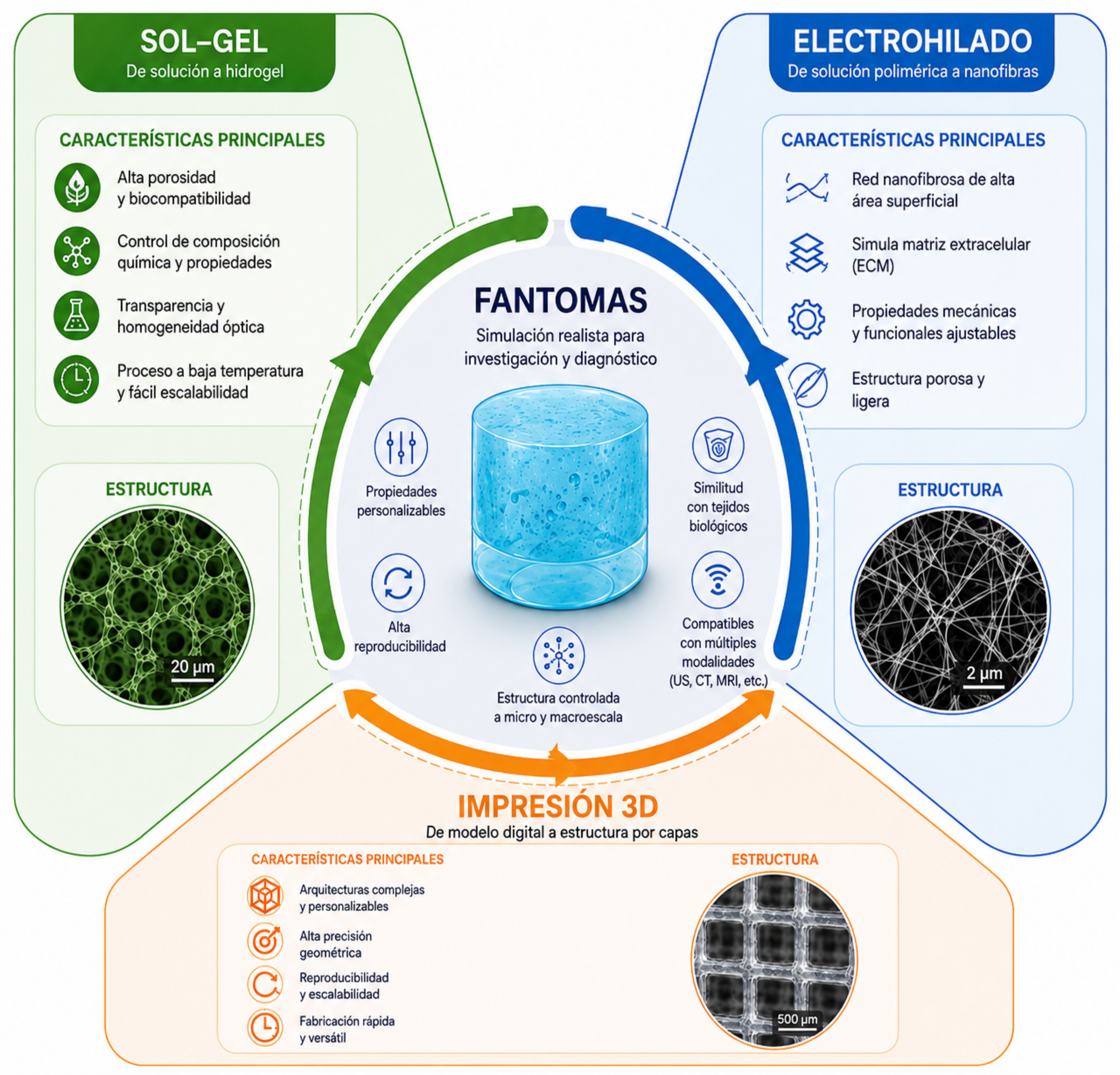
Los avances recientes en la preparación de hidrogeles han impulsado el diseño de fantomas biomédicos capaces de simular tejidos blandos y facilitar el estudio de propiedades biológicas, en particular la densidad aparente y efectiva. Estos avances permiten la calibración de equipos de imagenología, la validación de dispositivos médicos y el entrenamiento de personal clínico. La presente revisión bibliográfica de alcance, basada en el análisis sistemático de la literatura publicada entre 2020 y 2026 en las bases de datos Scopus, Web of Science, PubMed y Google Scholar, identifica y compara tres métodos de fabricación de fantomas ampliamente utilizados por su versatilidad operacional: métodos químicos de reticulación basados en procesos sol-gel, técnicas de impresión 3D y procesos de electrohilado. Cada método presenta ventajas específicas. Los métodos químicos destacan por su sencillez y estabilidad; la impresión 3D aporta control geométrico y reproducibilidad, posibilitando la creación de arquitecturas complejas que influyen directamente en la densidad volumétrica; mientras que el electrohilado permite crear micro y nanoestructuras fibrosas que imitan la matriz extracelular y ofrece un control preciso de la densidad a través del diámetro, la orientación y la fracción de vacíos entre las fibras. La evidencia disponible indica que la convergencia de los métodos de impresión 3D y electrohilado en enfoques híbridos representa una frontera tecnológica prometedora para superar las limitaciones individuales de cada técnica, lo que permite la fabricación de fantomas con fidelidad anatómica a escala macroscópica y mimetismo estructural a escala nanométrica. Los hidrogeles constituyen uno de los materiales más ampliamente utilizados en la fabricación de fantomas debido a su reproducibilidad, su simplicidad y su capacidad de incorporar partículas que mejoran la simulación de tejidos biológicos. Polímeros naturales como el quitosano, el alginato y el alcohol polivinílico permiten ajustar la densidad mediante la concentración polimérica, el grado de reticulación y la incorporación de agentes de relleno. La medición de la densidad en los hidrogeles es esencial para evaluar su interacción con fluidos biológicos y optimizar su rendimiento en aplicaciones como la terapia celular. Se examinan los materiales empleados, las propiedades ajustables, los métodos de caracterización y las aplicaciones clínicas de estos sistemas. Técnicas no invasivas, como la bioimpedancia eléctrica, la tomografía por ultrasonido y el ultrasonido inducido por láser, emplean hidrogeles para calibrar densidades tisulares, lo que mejora la calidad de las imágenes y amplía sus aplicaciones clínicas. En conjunto, estos avances consolidan a los hidrogeles como herramientas clave para el desarrollo de fantomas biomédicos más realistas y para su futura aplicación clínica. Como principal contribución, el trabajo propone un marco comparativo que orienta la selección de la tecnología según la aplicación biomédica, destacando la convergencia de la impresión 3D y el electrohilado como un enfoque híbrido prometedor. Adicionalmente, se identifican los principales desafíos en materia de estandarización, resolución multiescala y multifuncionalidad, y se delinean las perspectivas futuras en el desarrollo de materiales inteligentes y estrategias de fabricación híbrida.
Descargas
Referencias
[1] M. Higgins, S. Leung, and N. Radacsi, “3D printing surgical phantoms and their role in the visualization of medical procedures,” Annals of 3D Printed Medicine, vol. 6, p. 100057, 2022. doi: 10.1016/j.stlm.2022.100057. DOI: https://doi.org/10.1016/j.stlm.2022.100057
[2] C. K. McGarry, L. J. Grattan, A. M. Ivory, F. Leek, G. P. Liney, Y. Liu, P. Miloro, R. Rai, A. P. Robinson, A. J. Shih, B. Zeqiri, and C. H. Clark, “Tissue mimicking materials for imaging and therapy phantoms: a review,” Phys. Med. Biol., vol. 65, p. 23TR01, 2020. doi: 10.1088/1361-6560/abbd17. DOI: https://doi.org/10.1088/1361-6560/abbd17
[3] S. A. Armstrong, R. Jafary, J. S. Forsythe, and S. D. Gregory, “Tissue-Mimicking Materials for Ultrasound-Guided Needle Intervention Phantoms: A Comprehensive Review,” Ultrasound in Medicine & Biology, vol. 49, no. 1, pp. 18–30, 2023. doi: 10.1016/j.ultrasmedbio.2022.07.016. DOI: https://doi.org/10.1016/j.ultrasmedbio.2022.07.016
[4] J. R. Cook, R. R. Bouchard, and S. Y. Emelianov, “Tissue-mimicking phantoms for photoacoustic and ultrasonic imaging,” Biomed. Opt. Express, vol. 2, pp. 3193–3206, 2011. doi: 10.1364/BOE.2.003193. DOI: https://doi.org/10.1364/BOE.2.003193
[5] A. Tejo-Otero, F. Fenollosa-Artés, I. Achaerandio, S. Rey-Vinolas, I. Buj-Corral, M. A. Mateos-Timoneda, and E. Engel, “Soft-Tissue-Mimicking Using Hydrogels for the Development of Phantoms,” Gels, vol. 8, p. 40, 2022. doi: 10.3390/gels8010040. DOI: https://doi.org/10.3390/gels8010040
[6] L. Wei, S. Wang, M. Shan, Y. Li, Y. Wang, F. Wang, L. Wang, and J. Mao, “Conductive fibers for biomedical applications,” Bioactive Materials, vol. 22, pp. 343–364, 2023. doi: 10.1016/j.bioactmat.2022.10.014. DOI: https://doi.org/10.1016/j.bioactmat.2022.10.014
[7] K. J. Juárez-Navarro, V. Guarino, and M. A. Álvarez-Pérez, “Converging Electrospinning and 3D-Printing Technologies: From Innovative Design for Tissue Engineering to Global Patent Trends and Technology Transfer,” Fibers, vol. 13, no. 6, p. 83, 2025. doi: 10.3390/fib13060083. DOI: https://doi.org/10.3390/fib13060083
[8] M. Kulmaganbetov, “Advancements in Retinal Tissue-Mimicking Optical Coherence Tomography Phantoms: Materials, Properties, and Applications,” BioChem, vol. 5, no. 2, p. 6, 2025. doi: 10.3390/biochem5020006. DOI: https://doi.org/10.3390/biochem5020006
[9] M. Zhang, S. Xu, R. Wang, Y. Che, C. Han, W. Feng, C. Wang, and W. Zhao, “Electrospun nanofiber/hydrogel composite materials and their tissue engineering applications,” Journal of Materials Science & Technology, vol. 162, pp. 157–178, 2023. doi: 10.1016/j.jmst.2023.04.015. DOI: https://doi.org/10.1016/j.jmst.2023.04.015
[10] M. Mabrouk, H. H. Beherei, and D. B. Das, “Recent progress in the fabrication techniques of 3D scaffolds for tissue engineering,” Mater. Sci. Eng. C Mater. Biol. Appl., vol. 110, p. 110716, 2020. doi: 10.1016/j.msec.2020.110716. DOI: https://doi.org/10.1016/j.msec.2020.110716
[11] M. R. Ghoto, B. H. Daubert, D. Parra-Cervantes, A. J. Hemmerla, B. D. Ulery, D. W. Hairston, C. G. Sinks, S. Young, and C. S. O’Bryan, “3D-Printing Soft Tissue Phantom Models from Photo-Crosslinkable Poly (vinyl alcohol) Methacrylate,” Materialia, vol. 44, p. 102592, 2025. doi: 10.1016/j.mtla.2025.102592.
[12] M. Zhang, J. Xing, Y. Zhong, T. Zhang, X. Liu, and D. Xing, “Advanced function, design and application of skin substitutes for skin regeneration,” Materials Today Bio, vol. 24, p. 100918, 2024. doi: 10.1016/j.mtbio.2023.100918. DOI: https://doi.org/10.1016/j.mtbio.2023.100918
[13] Y. Li, X. Li, Z. Liu, Y. Wang, and T. Jiao, “Recent Progress in the Strategies and Applications of Electrospinning Electroactive Tissue Engineering Scaffolds,” ACS Biomater. Sci. Eng., vol. 11, no. 6, pp. 3182–3200, 2025. doi: 10.1021/acsbiomaterials.5c00142. DOI: https://doi.org/10.1021/acsbiomaterials.5c00142
[14] S. Pawel, “Electroconductive scaffolds for tissue engineering applications,” J. Appl. Mater. Interfaces, vol. 8, p. 20, pp. 2047–4830, 2020. doi: 10.1039/D0BM01176B. DOI: https://doi.org/10.1039/D0BM01176B
[15] T. Khan, G. Vadivel, K. Ayyasamy, G. Murugesan, and T. A. Sebaey, “Advances in Conductive Biomaterials for Cardiac Tissue Engineering: Design, Fabrication, and Functional Integration,” Polymers, vol. 17, no. 5, p. 620, 2025. doi: 10.3390/polym17050620. DOI: https://doi.org/10.3390/polym17050620
[16] J. Wu, F. Yu, M. Shao, T. Zhang, W. Lu, X. Chen, Y. Wang, and Y. Guo, “Electrospun Nanofiber Scaffold for Skin Tissue Engineering: A Review,” ACS Applied Bio Materials, vol. 7, no. 6, pp. 3556–3567, 2024. doi: 10.1021/acsabm.4c00318. DOI: https://doi.org/10.1021/acsabm.4c00318
[17] X. Ma, M. Xu, X. Cui, J. Yin, and Q. Wu, “Hybrid 3D Bioprinting of Sustainable Biomaterials for Advanced Multiscale Tissue Engineering,” Small, vol. 22, no. 16, p. 2408947, 2026. doi: 10.1002/smll.202408947. DOI: https://doi.org/10.1002/smll.202408947
[18] X. Gu, Z. Shu, X. Zheng, S. Wei, M. Ma, H. He, Y. Shi, X. Gong, S. Chen, and X. Wang, “A novel CT-responsive hydrogel for the construction of an organ simulation phantom for the repeatability and stability study of radiomic features,” J. Mater. Chem. B, vol. 11, no. 46, pp. 11073–11081, 2023. doi: 10.1039/d3tb01706k. DOI: https://doi.org/10.1039/D3TB01706K
[19] J. Yin, M. Li, G. Dai, et al., “3D Printed Multi-material Medical Phantoms for Needle-tissue Interaction Modelling of Heterogeneous Structures,” J. Bionic Eng., vol. 18, pp. 346–360, 2021. doi: 10.1007/s42235-021-0031-1. DOI: https://doi.org/10.1007/s42235-021-0031-1
[20] A. Jawli, A. Wadhhah, N. Ghulam, and H. Zhihong, “Tissue-Mimicking Material Fabrication and Properties for Multiparametric Ultrasound Phantoms: A Systematic Review,” Bioengineering, vol. 11, no. 6, p. 620, 2024. doi: 10.3390/bioengineering11060620. DOI: https://doi.org/10.3390/bioengineering11060620
[21] A. Bublex, A. Montalibet, B. Massot, and C. Gehin, “Review of Phantoms for Mimicking the Electrical Properties and Mechanical Strength of Living Tissue,” IRBM, vol. 46, no. 5, p. 100904, 2025. doi: 10.1016/j.irbm.2025.100904. DOI: https://doi.org/10.1016/j.irbm.2025.100904
[22] C. J. Brinker and G. W. Scherer, Sol-Gel Science: The Physics and Chemistry of Sol-Gel Processing. Academic Press, 1990; J. Livage, “Sol-gel chemistry of transition metal oxides,” Curr. Opin. Solid State Mater. Sci., vol. 3, no. 5, pp. 231–235, 1998.
[23] H. Matsuyama, M. Teramoto, and H. Urano, “Photopolymerization of acrylamide in sol-gel derived silica,” J. Sol-Gel Sci. Technol., vol. 25, no. 1, pp. 75–81, 2002.
[24] J. Li and D. J. Mooney, “Designing Hydrogels for Controlled Drug Delivery,” Nat. Rev. Mater., vol. 1, p. 16071, 2016. DOI: https://doi.org/10.1038/natrevmats.2016.71
[25] E. In, “Development of Polymer Based Gels for Multimodal Medical Imaging Phantoms,” Ph.D. dissertation, Univ. of Toronto, 2016.
[26] M. Matsusaki, et al., “Fabrication of Perfusable Pseudo Blood Vessels by Controlling Sol Gel Transition of Gellan Gum Templates,” ACS Biomater. Sci. Eng., vol. 5, no. 11, pp. 5637–5643, 2019. doi: 10.1021/acsbiomaterials.8b01272.
[27] A. Karadeniz-Yildirim and H. Tanyildizi-Kokkulunk, “Investigation of the tissue equivalence of typical 3D-printing materials for application in internal dosimetry using monte carlo simulations,” Phys. Eng. Sci. Med., vol. 48, pp. 665–673, 2025. doi: 10.1007/s13246-025-01532-2. DOI: https://doi.org/10.1007/s13246-025-01532-2
[28] C. Echalier, L. Raimond, M. Simon, et al., “Hybrid hydrogels: artificial extracellular matrices for regenerative therapies,” J. Pept. Sci., vol. 28, no. S3, p. P197, 2022.
[29] C. S. O’Bryan, et al., “3D printing soft tissue phantom models from photo crosslinkable poly(vinyl alcohol) methacrylate,” Materialia, vol. 44, p. 102592, 2025. DOI: https://doi.org/10.1016/j.mtla.2025.102592
[30] K. Zöller and A. To, “Biomedical applications of functional hydrogels: Innovative developments, relevant clinical trials and advanced products,” Biomaterials, vol. 312, p. 122718, 2025. doi: 10.1016/j.biomaterials.2024.122718. DOI: https://doi.org/10.1016/j.biomaterials.2024.122718
[31] M. Gonçalves, Y. T. Ong, S. Baek, S. Kim, and J. W. Kim, “Smart Sol Gel Hydrogels for Disease Targeted Therapeutics: Molecular Design and Biomedical Translation,” Adv. Funct. Mater., p. 231972, 2026. DOI: https://doi.org/10.1002/adfm.202531972
[32] Z. Zhao, Z. Qin, T. Zhao, Y. Li, Z. Hou, H. Hu, X. Su, and Y. Gao, “Crosslinked Biodegradable Hybrid Hydrogels Based on Poly(ethylene glycol) and Gelatin for Drug Controlled Release,” Molecules, vol. 29, no. 20, p. 4952, 2024. doi: 10.3390/molecules29204952. DOI: https://doi.org/10.3390/molecules29204952
[33] K. Yue, G. Trujillo-de Santiago, M. M. Alvarez, A. Tamayol, N. Annabi, and A. Khademhosseini, “Synthesis, properties, and biomedical applications of gelatin methacryloyl (GelMA) hydrogels,” Biomaterials, vol. 73, pp. 254–271, 2015. doi: 10.1016/j.biomaterials.2015.08.045.
[34] J. H. Lee, “Injectable hydrogels delivering therapeutic agents for disease treatment and tissue engineering,” Biomater. Res., vol. 22, p. 27, 2018. doi: 10.1186/s40824-018-0138-6. DOI: https://doi.org/10.1186/s40824-018-0138-6
[35] W. Song, J. Ko, Y. H. Choi, and N. S. Hwang, “Recent advancements in enzyme-mediated crosslinkable hydrogels: In vivo-mimicking strategies,” APL Bioeng., vol. 5, no. 2, p. 021502, 2021. doi: 10.1063/5.0037793. DOI: https://doi.org/10.1063/5.0037793
[36] M. Okawa, A. Tanabe, S. Ohta, S. Nagatoishi, K. Tsumoto, and T. Ito, “Extracellular matrix-inspired hydrogel of hyaluronan and gelatin crosslinked via a Link module with a transglutaminase reactive sequence,” Commun. Mater., vol. 3, p. 81, 2022. doi: 10.1038/s43246-022-00249-9. DOI: https://doi.org/10.1038/s43246-022-00309-4
[37] K. J. M. Surry, H. J. B. Austin, A. Fenster, and T. M. Peters, “Poly(vinyl alcohol) cryogel phantoms for use in ultrasound and MR imaging,” Phys. Med. Biol., vol. 49, no. 24, pp. 5529–5546, 2004. doi: 10.1088/0031-9155/49/24/009. DOI: https://doi.org/10.1088/0031-9155/49/24/009
[38] M. Matsusaki, H. Ikeguchi, C. Kubo, H. Sato, Y. Kuramochi, and D. Takagi, “Fabrication of Perfusable Pseudo Blood Vessels by Controlling Sol-Gel Transition of Gellan Gum Templates,” ACS Biomater. Sci. Eng., vol. 5, no. 11, pp. 5637–5643, 2019. doi: 10.1021/acsbiomaterials.8b01272. DOI: https://doi.org/10.1021/acsbiomaterials.8b01272
[39] R. A. A. Muzzarelli, M. El Mehtedi, C. Bottegoni, A. Aquili, and A. Gigante, “Genipin-Crosslinked Chitosan Gels and Scaffolds for Tissue Engineering and Regeneration of Cartilage and Bone,” Mar. Drugs, vol. 13, no. 12, pp. 7314–7338, 2015. doi: 10.3390/md13127068. DOI: https://doi.org/10.3390/md13127068
[40] Á. Castro-María, J. P. Fernández-Blázquez, and J. Patterson, “Genipin-Crosslinked Gelatin Hydrogels with Controlled Molecular Weight: A Strategy to Balance Processability and Performance,” Gels, vol. 11, no. 12, p. 980, 2025. doi: 10.3390/gels11120980. DOI: https://doi.org/10.3390/gels11120980
[41] Á. Rodríguez-González, L. M. Delgado, and R. A. Pérez, “Self-setting silica-gelatin hybrid 3D printed scaffolds with tunable composition and enhanced physicochemical and biological properties,” J. Sol-Gel Sci. Technol., vol. 117, no. 3, 2026. doi: 10.1007/s10971-025-06678-9.
[42] A. Jawli, G. Nabi, and Z. Huang, “A Polyvinyl Alcohol (PVA)-Based Phantom for Prostate Cancer Detection Using Multiparametric Ultrasound: A Validation Study,” Bioengineering, vol. 11, no. 11, p. 1052, 2024. doi: 10.3390/bioengineering11111052. DOI: https://doi.org/10.3390/bioengineering11111052
[43] W. D’Souza, E. Madsen, O. Unal, K. Vigen, G. Frank, and B. Thomadsen, “Tissue mimicking materials for a multi-imaging modality prostate phantom,” Med. Phys., vol. 28, no. 4, pp. 688–700, 2021. doi: 10.1118/1.1354998. DOI: https://doi.org/10.1118/1.1354998
[44] Á. Rodríguez-González, L. M. Delgado, and R. A. Pérez, “Self-setting silica-gelatin hybrid 3D printed scaffolds with tunable composition and enhanced physicochemical and biological properties,” J. Sol-Gel Sci. Technol., vol. 117, no. 3, 2026. doi: 10.1007/s10971-025-06678-9. DOI: https://doi.org/10.1007/s10971-025-07022-x
[45] E. In, “Development of Polymer Based Gels for Multimodal Medical Imaging Phantoms,” Ph.D. dissertation, Univ. of Toronto, 2016.
[46] K. Haraguchi and T. Takehisa, “Nanocomposite hydrogels: A unique organic–inorganic network structure with extraordinary mechanical, optical, and swelling/de-swelling properties,” Adv. Mater., vol. 14, no. 16, pp. 1120–1124, 2002. doi: 10.1002/1521-4095(20020816)14:16<1120::AID-ADMA1120>3.0.CO;2-9. DOI: https://doi.org/10.1002/1521-4095(20020816)14:16<1120::AID-ADMA1120>3.0.CO;2-9
[47] H. Fan, J. Wang, Q. Zhang, Z. Jin, G. Tsuji, Y. Zhang, and H. Gao, “Supramolecular hydrogel with high mechanical strength and self-healing properties,” Nat. Commun., vol. 6, p. 7418, 2015. doi: 10.1038/ncomms8418. DOI: https://doi.org/10.1038/ncomms8418
[48] T. Mazaki, Y. Shiozaki, K. Yamane, A. Yoshida, M. Nakamura, Y. Yoshida, D. Zhou, T. Kitajima, M. Tanaka, Y. Ito, T. Ozaki, and A. Matsukawa, “A novel, visible light-induced, rapidly cross-linkable gelatin scaffold for osteochondral tissue engineering,” Sci. Rep., vol. 4, p. 4457, 2014. doi: 10.1038/srep04457. DOI: https://doi.org/10.1038/srep04457
[49] J. L. Drury, R. G. Dennis, and D. J. Mooney, “The tensile properties of alginate hydrogels,” Biomaterials, vol. 25, no. 16, pp. 3187–3199, 2004. doi: 10.1016/j.biomaterials.2003.10.002. DOI: https://doi.org/10.1016/j.biomaterials.2003.10.002
[50] K. Yue, G. Trujillo-de Santiago, M. M. Alvarez, A. Tamayol, N. Annabi, and A. Khademhosseini, “Synthesis, properties, and biomedical applications of gelatin methacryloyl (GelMA) hydrogels,” Biomaterials, vol. 73, pp. 254–271, 2015. doi: 10.1016/j.biomaterials.2015.08.045. DOI: https://doi.org/10.1016/j.biomaterials.2015.08.045
[51] A. Filippou and C. Damianou, “Agar-based phantom for evaluating targeting of high-intensity focused ultrasound systems for breast ablation,” J. Med. Phys., vol. 49, no. 3, pp. 343–355, 2024. doi: 10.4103/jmp.jmp_52_24. DOI: https://doi.org/10.4103/jmp.jmp_52_24
[52] X. Liang, H.-J. Zhong, H. Ding, B. Yu, X. Ma, X. Liu, C.-M. Chong, and J. He, “Polyvinyl Alcohol (PVA)-Based Hydrogels: Recent Progress in Fabrication, Properties, and Multifunctional Applications,” Polymers, vol. 16, no. 19, p. 2755, 2024. doi: 10.3390/polym16192755. DOI: https://doi.org/10.3390/polym16192755
[53] E. Barrett-Catton, M. L. Ross, and P. Asuri, “Multifunctional Hydrogel Nanocomposites for Biomedical Applications,” Polymers, vol. 13, no. 6, p. 856, 2021. doi: 10.3390/polym13060856. DOI: https://doi.org/10.3390/polym13060856
[54] G. Baishya, B. Parasar, M. Limboo, R. Kumar, A. Dutta, A. Hussain, M. M. Phukan, and D. Saikia, “Advancements in nanocomposite hydrogels: A comprehensive review of biomedical applications,” Discover Mater., vol. 4, p. 40, 2024. doi: 10.1007/s44154-024-00040-9. DOI: https://doi.org/10.1007/s43939-024-00111-8
[55] X. Zhang, W. Yang, H. Zhang, M. Xie, and X. Duan, “PEDOT:PSS: From conductive polymers to sensors,” Nanotechnol. Precis. Eng., vol. 4, no. 4, p. 045004, 2021. doi: 10.1063/10.0006866. DOI: https://doi.org/10.1063/10.0006866
[56] J. Li, D. Mo, J. Hu, S. Wang, J. Gong, Y. Huang, Z. Li, Z. Yuan, and M. Xu, “PEDOT:PSS-based bioelectronics for brain monitoring and modulation,” Microsyst. Nanoeng., vol. 11, p. 87, 2025. doi: 10.1038/s41378-025-00687-9. DOI: https://doi.org/10.1038/s41378-025-00948-w
[57] H. Wang, X. Sun, Y. Wang, K. Li, J. Wang, X. Dai, B. Chen, D. Chong, L. Zhang, and J. Yan, “Acid enhanced zipping effect to densify MWCNT packing for multifunctional MWCNT films with ultra-high electrical conductivity,” Nat. Commun., vol. 14, p. 380, 2023. doi: 10.1038/s41467-023-380. DOI: https://doi.org/10.1038/s41467-023-36082-2
[58] A. P. Rath, S. G. Krishnan, and K. Kanny, “Studies on (polytrimethylene terephthalate)/graphene oxide/f-MWCNT hybrid nanocomposites,” Discover Nano, vol. 19, p. 21, 2024. doi: 10.1007/s44154-024-00021-9. DOI: https://doi.org/10.1186/s11671-024-03966-1
[59] M. Rai, A. Yadav, and A. Gade, “Silver nanoparticles as a new generation of antimicrobials,” Biotechnol. Adv., vol. 27, no. 1, pp. 76–83, 2009. doi: 10.1016/j.biotechadv.2008.09.002. DOI: https://doi.org/10.1016/j.biotechadv.2008.09.002
[60] E. C. Dreaden, A. M. Alkilany, X. Huang, C. J. Murphy, and M. A. El-Sayed, “The golden age: gold nanoparticles for biomedicine,” Chem. Soc. Rev., vol. 41, no. 7, pp. 2740–2779, 2012. doi: 10.1039/C1CS15237H. DOI: https://doi.org/10.1039/C1CS15237H
[61] H. Lin, Y. Chen, and J. Shi, “MXenes for next-generation biomedical applications,” Adv. Drug Deliv. Rev., vol. 138, pp. 196–213, 2018. doi: 10.1016/j.addr.2018.01.002. DOI: https://doi.org/10.1016/j.addr.2018.01.002
[62] B. Shao, H. Lin, and Y. Chen, “MXene nanomaterials in biomedicine: applications and prospects,” Adv. Mater., vol. 32, no. 23, p. 1905677, 2020. doi: 10.1002/adma.201905677.
[63] G. G. Flores-Rojas, et al., “Electrospun scaffolds for tissue engineering: A review,” Macromol., vol. 3, no. 3, p. 31, 2021. doi: 10.3390/macromol3030031. DOI: https://doi.org/10.3390/macromol3030031
[64] L. Muthukrishnan, “An overview on electrospinning and its advancement toward hard and soft tissue engineering applications,” Colloid Polym. Sci., vol. 300, no. 8, pp. 875–901, 2022. doi: 10.1007/s00396-022-04997-9. DOI: https://doi.org/10.1007/s00396-022-04997-9
[65] A. Kumara and D. Sheet, “Method for Developing Tissue-mimicking Ultrasound Imaging Phantoms of Organs using Polyvinyl Alcohol and 3-D Printed Moulds,” Ultrasound Med. Biol., vol. 52, pp. 401–410, 2026. doi: 10.1016/j.ultrasmedbio.2025.10.006. DOI: https://doi.org/10.1016/j.ultrasmedbio.2025.10.006
[66] A. Antoniou and C. Damianou, “MR relaxation properties of tissue-mimicking phantoms,” Ultrasonics, vol. 119, p. 106600, 2022. doi: 10.1016/j.ultras.2021.106600. DOI: https://doi.org/10.1016/j.ultras.2021.106600
[67] A. Antoniou, L. Georgiou, T. Christodoulou, N. Panayiotou, C. Ioannides, N. Zamboglou, and C. Damianou, “MR relaxation times of agar-based tissue-mimicking phantoms,” J. Appl. Clin. Med. Phys., vol. 23, no. 5, p. e13533, 2022. doi: 10.1002/acm2.13533. DOI: https://doi.org/10.1002/acm2.13533
Publicado
Declaración de disponibilidad de datos
a solicitud de los interesados
Número
Sección
Categorías
Licencia
Derechos de autor 2026 Eduardo Martínez-Ramírez, David Gasca-Figueroa, Abisaí Jaime Reséndiz-Barrón, Rosa María Quispe-Siccha, Gerardo Gutiérrez-Juárez, Francisco Javier García-Rodríguez

Esta obra está bajo una licencia internacional Creative Commons Atribución 4.0.
Los autores/as que publiquen en esta revista aceptan las siguientes condiciones:
- Los autores/as conservan los derechos de autor y ceden a la revista el derecho de la primera publicación, con el trabajo registrado con la licencia de atribución de Creative Commons 4.0, que permite a terceros utilizar lo publicado siempre que mencionen la autoría del trabajo y a la primera publicación en esta revista.
- Los autores/as pueden realizar otros acuerdos contractuales independientes y adicionales para la distribución no exclusiva de la versión del artículo publicado en esta revista (p. ej., incluirlo en un repositorio institucional o publicarlo en un libro) siempre que indiquen claramente que el trabajo se publicó por primera vez en esta revista.
- Se permite y recomienda a los autores/as a compartir su trabajo en línea (por ejemplo: en repositorios institucionales o páginas web personales) antes y durante el proceso de envío del manuscrito, ya que puede conducir a intercambios productivos, a una mayor y más rápida citación del trabajo publicado (vea The Effect of Open Access).
Cómo citar