Hydrogels for soft-tissue phantoms: From Sol-Gel Methods to Hybrid Frontiers in 3D Printing and Electrospinning
DOI:
https://doi.org/10.37636/recit.v9n3e470Keywords:
Hydrogels, Tissues Phantoms, Sol-gel, 3D printing, Electrospinning, Tissue engineering, Biomimetic materialsAbstract
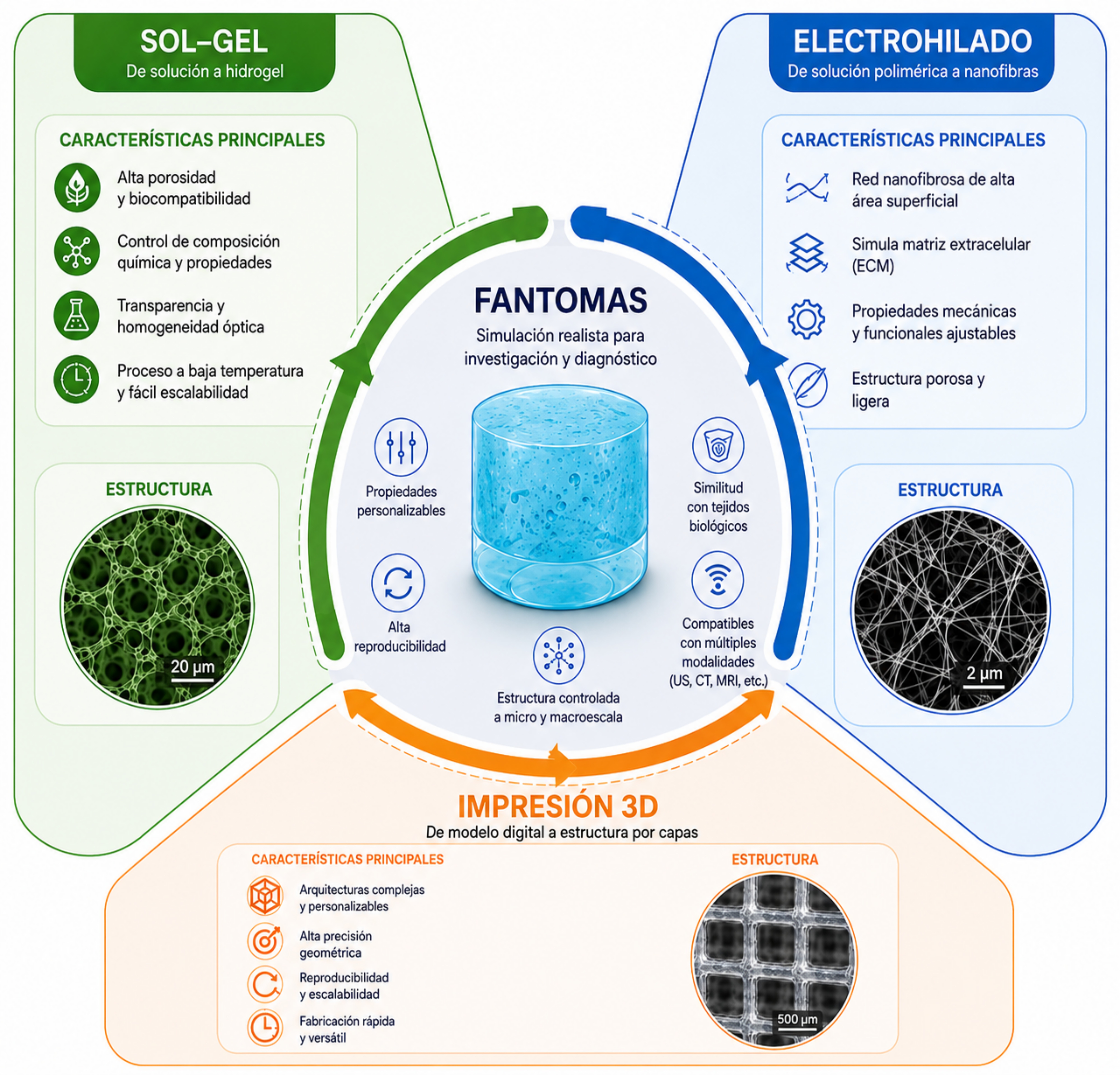
Recent advances in hydrogel preparation have driven the design of biomedical phantoms capable of simulating soft tissues and facilitating the study of biological properties, particularly apparent and effective density. These advances enable the calibration of imaging equipment, the validation of medical devices, and the training of clinical personnel. This scoping review, based on a systematic analysis of the literature published between 2020 and 2026 in Scopus, Web of Science, PubMed, and Google Scholar, identifies and compares three phantom fabrication methods widely used due to their operational versatility: chemical crosslinking methods based on sol-gel processes, 3D printing techniques, and electrospinning processes. Each method has specific advantages. Chemical methods stand out for their simplicity and stability; 3D printing provides geometric control and reproducibility, enabling the creation of complex architectures that directly influence volumetric density; while electrospinning allows the creation of fibrous micro- and nanostructures that mimic the extracellular matrix, offering precise density control through fiber diameter, orientation, and void fraction. Current evidence suggests that hybrid approaches combining 3D printing and electrospinning constitute a promising technological frontier, as they overcome the limitations of each individual technique and allow the fabrication of phantoms that integrate macroscopic anatomical fidelity with nanoscale structural mimicry. Hydrogels constitute the most widely used material in phantom fabrication due to their reproducibility, simplicity, and ability to incorporate particles that enhance the simulation of biological tissues. Natural polymers such as chitosan, alginate, and polyvinyl alcohol allow density adjustment through polymer concentration, degree of crosslinking, and the incorporation of filler agents. Density measurement in hydrogels is essential to evaluate their interaction with biological fluids and optimize their performance in applications such as cell therapy. The materials used, tunable properties, characterization methods, and clinical applications of these systems are examined. Noninvasive techniques such as electrical bioimpedance, ultrasound tomography, and laser induced ultrasound employ hydrogels to calibrate tissue densities, thereby improving image quality and expanding their clinical applications. Together, these advances consolidate hydrogels as key tools for the development of more realistic biomedical phantoms and for their future clinical application. As its main contribution, the work proposes a comparative framework that guides the selection of technology according to the biomedical application, highlighting the convergence of 3D printing and electrospinning as a promising hybrid approach. Additionally, the main challenges in terms of standardization, multiscale resolution, and multifunctionality are identified, and future perspectives are outlined for the development of smart materials and hybrid manufacturing strategies.
Downloads
References
[1] M. Higgins, S. Leung, and N. Radacsi, “3D printing surgical phantoms and their role in the visualization of medical procedures,” Annals of 3D Printed Medicine, vol. 6, p. 100057, 2022. doi: 10.1016/j.stlm.2022.100057. DOI: https://doi.org/10.1016/j.stlm.2022.100057
[2] C. K. McGarry, L. J. Grattan, A. M. Ivory, F. Leek, G. P. Liney, Y. Liu, P. Miloro, R. Rai, A. P. Robinson, A. J. Shih, B. Zeqiri, and C. H. Clark, “Tissue mimicking materials for imaging and therapy phantoms: a review,” Phys. Med. Biol., vol. 65, p. 23TR01, 2020. doi: 10.1088/1361-6560/abbd17. DOI: https://doi.org/10.1088/1361-6560/abbd17
[3] S. A. Armstrong, R. Jafary, J. S. Forsythe, and S. D. Gregory, “Tissue-Mimicking Materials for Ultrasound-Guided Needle Intervention Phantoms: A Comprehensive Review,” Ultrasound in Medicine & Biology, vol. 49, no. 1, pp. 18–30, 2023. doi: 10.1016/j.ultrasmedbio.2022.07.016. DOI: https://doi.org/10.1016/j.ultrasmedbio.2022.07.016
[4] J. R. Cook, R. R. Bouchard, and S. Y. Emelianov, “Tissue-mimicking phantoms for photoacoustic and ultrasonic imaging,” Biomed. Opt. Express, vol. 2, pp. 3193–3206, 2011. doi: 10.1364/BOE.2.003193. DOI: https://doi.org/10.1364/BOE.2.003193
[5] A. Tejo-Otero, F. Fenollosa-Artés, I. Achaerandio, S. Rey-Vinolas, I. Buj-Corral, M. A. Mateos-Timoneda, and E. Engel, “Soft-Tissue-Mimicking Using Hydrogels for the Development of Phantoms,” Gels, vol. 8, p. 40, 2022. doi: 10.3390/gels8010040. DOI: https://doi.org/10.3390/gels8010040
[6] L. Wei, S. Wang, M. Shan, Y. Li, Y. Wang, F. Wang, L. Wang, and J. Mao, “Conductive fibers for biomedical applications,” Bioactive Materials, vol. 22, pp. 343–364, 2023. doi: 10.1016/j.bioactmat.2022.10.014. DOI: https://doi.org/10.1016/j.bioactmat.2022.10.014
[7] K. J. Juárez-Navarro, V. Guarino, and M. A. Álvarez-Pérez, “Converging Electrospinning and 3D-Printing Technologies: From Innovative Design for Tissue Engineering to Global Patent Trends and Technology Transfer,” Fibers, vol. 13, no. 6, p. 83, 2025. doi: 10.3390/fib13060083. DOI: https://doi.org/10.3390/fib13060083
[8] M. Kulmaganbetov, “Advancements in Retinal Tissue-Mimicking Optical Coherence Tomography Phantoms: Materials, Properties, and Applications,” BioChem, vol. 5, no. 2, p. 6, 2025. doi: 10.3390/biochem5020006. DOI: https://doi.org/10.3390/biochem5020006
[9] M. Zhang, S. Xu, R. Wang, Y. Che, C. Han, W. Feng, C. Wang, and W. Zhao, “Electrospun nanofiber/hydrogel composite materials and their tissue engineering applications,” Journal of Materials Science & Technology, vol. 162, pp. 157–178, 2023. doi: 10.1016/j.jmst.2023.04.015. DOI: https://doi.org/10.1016/j.jmst.2023.04.015
[10] M. Mabrouk, H. H. Beherei, and D. B. Das, “Recent progress in the fabrication techniques of 3D scaffolds for tissue engineering,” Mater. Sci. Eng. C Mater. Biol. Appl., vol. 110, p. 110716, 2020. doi: 10.1016/j.msec.2020.110716. DOI: https://doi.org/10.1016/j.msec.2020.110716
[11] M. R. Ghoto, B. H. Daubert, D. Parra-Cervantes, A. J. Hemmerla, B. D. Ulery, D. W. Hairston, C. G. Sinks, S. Young, and C. S. O’Bryan, “3D-Printing Soft Tissue Phantom Models from Photo-Crosslinkable Poly (vinyl alcohol) Methacrylate,” Materialia, vol. 44, p. 102592, 2025. doi: 10.1016/j.mtla.2025.102592.
[12] M. Zhang, J. Xing, Y. Zhong, T. Zhang, X. Liu, and D. Xing, “Advanced function, design and application of skin substitutes for skin regeneration,” Materials Today Bio, vol. 24, p. 100918, 2024. doi: 10.1016/j.mtbio.2023.100918. DOI: https://doi.org/10.1016/j.mtbio.2023.100918
[13] Y. Li, X. Li, Z. Liu, Y. Wang, and T. Jiao, “Recent Progress in the Strategies and Applications of Electrospinning Electroactive Tissue Engineering Scaffolds,” ACS Biomater. Sci. Eng., vol. 11, no. 6, pp. 3182–3200, 2025. doi: 10.1021/acsbiomaterials.5c00142. DOI: https://doi.org/10.1021/acsbiomaterials.5c00142
[14] S. Pawel, “Electroconductive scaffolds for tissue engineering applications,” J. Appl. Mater. Interfaces, vol. 8, p. 20, pp. 2047–4830, 2020. doi: 10.1039/D0BM01176B. DOI: https://doi.org/10.1039/D0BM01176B
[15] T. Khan, G. Vadivel, K. Ayyasamy, G. Murugesan, and T. A. Sebaey, “Advances in Conductive Biomaterials for Cardiac Tissue Engineering: Design, Fabrication, and Functional Integration,” Polymers, vol. 17, no. 5, p. 620, 2025. doi: 10.3390/polym17050620. DOI: https://doi.org/10.3390/polym17050620
[16] J. Wu, F. Yu, M. Shao, T. Zhang, W. Lu, X. Chen, Y. Wang, and Y. Guo, “Electrospun Nanofiber Scaffold for Skin Tissue Engineering: A Review,” ACS Applied Bio Materials, vol. 7, no. 6, pp. 3556–3567, 2024. doi: 10.1021/acsabm.4c00318. DOI: https://doi.org/10.1021/acsabm.4c00318
[17] X. Ma, M. Xu, X. Cui, J. Yin, and Q. Wu, “Hybrid 3D Bioprinting of Sustainable Biomaterials for Advanced Multiscale Tissue Engineering,” Small, vol. 22, no. 16, p. 2408947, 2026. doi: 10.1002/smll.202408947. DOI: https://doi.org/10.1002/smll.202408947
[18] X. Gu, Z. Shu, X. Zheng, S. Wei, M. Ma, H. He, Y. Shi, X. Gong, S. Chen, and X. Wang, “A novel CT-responsive hydrogel for the construction of an organ simulation phantom for the repeatability and stability study of radiomic features,” J. Mater. Chem. B, vol. 11, no. 46, pp. 11073–11081, 2023. doi: 10.1039/d3tb01706k. DOI: https://doi.org/10.1039/D3TB01706K
[19] J. Yin, M. Li, G. Dai, et al., “3D Printed Multi-material Medical Phantoms for Needle-tissue Interaction Modelling of Heterogeneous Structures,” J. Bionic Eng., vol. 18, pp. 346–360, 2021. doi: 10.1007/s42235-021-0031-1. DOI: https://doi.org/10.1007/s42235-021-0031-1
[20] A. Jawli, A. Wadhhah, N. Ghulam, and H. Zhihong, “Tissue-Mimicking Material Fabrication and Properties for Multiparametric Ultrasound Phantoms: A Systematic Review,” Bioengineering, vol. 11, no. 6, p. 620, 2024. doi: 10.3390/bioengineering11060620. DOI: https://doi.org/10.3390/bioengineering11060620
[21] A. Bublex, A. Montalibet, B. Massot, and C. Gehin, “Review of Phantoms for Mimicking the Electrical Properties and Mechanical Strength of Living Tissue,” IRBM, vol. 46, no. 5, p. 100904, 2025. doi: 10.1016/j.irbm.2025.100904. DOI: https://doi.org/10.1016/j.irbm.2025.100904
[22] C. J. Brinker and G. W. Scherer, Sol-Gel Science: The Physics and Chemistry of Sol-Gel Processing. Academic Press, 1990; J. Livage, “Sol-gel chemistry of transition metal oxides,” Curr. Opin. Solid State Mater. Sci., vol. 3, no. 5, pp. 231–235, 1998.
[23] H. Matsuyama, M. Teramoto, and H. Urano, “Photopolymerization of acrylamide in sol-gel derived silica,” J. Sol-Gel Sci. Technol., vol. 25, no. 1, pp. 75–81, 2002.
[24] J. Li and D. J. Mooney, “Designing Hydrogels for Controlled Drug Delivery,” Nat. Rev. Mater., vol. 1, p. 16071, 2016. DOI: https://doi.org/10.1038/natrevmats.2016.71
[25] E. In, “Development of Polymer Based Gels for Multimodal Medical Imaging Phantoms,” Ph.D. dissertation, Univ. of Toronto, 2016.
[26] M. Matsusaki, et al., “Fabrication of Perfusable Pseudo Blood Vessels by Controlling Sol Gel Transition of Gellan Gum Templates,” ACS Biomater. Sci. Eng., vol. 5, no. 11, pp. 5637–5643, 2019. doi: 10.1021/acsbiomaterials.8b01272.
[27] A. Karadeniz-Yildirim and H. Tanyildizi-Kokkulunk, “Investigation of the tissue equivalence of typical 3D-printing materials for application in internal dosimetry using monte carlo simulations,” Phys. Eng. Sci. Med., vol. 48, pp. 665–673, 2025. doi: 10.1007/s13246-025-01532-2. DOI: https://doi.org/10.1007/s13246-025-01532-2
[28] C. Echalier, L. Raimond, M. Simon, et al., “Hybrid hydrogels: artificial extracellular matrices for regenerative therapies,” J. Pept. Sci., vol. 28, no. S3, p. P197, 2022.
[29] C. S. O’Bryan, et al., “3D printing soft tissue phantom models from photo crosslinkable poly(vinyl alcohol) methacrylate,” Materialia, vol. 44, p. 102592, 2025. DOI: https://doi.org/10.1016/j.mtla.2025.102592
[30] K. Zöller and A. To, “Biomedical applications of functional hydrogels: Innovative developments, relevant clinical trials and advanced products,” Biomaterials, vol. 312, p. 122718, 2025. doi: 10.1016/j.biomaterials.2024.122718. DOI: https://doi.org/10.1016/j.biomaterials.2024.122718
[31] M. Gonçalves, Y. T. Ong, S. Baek, S. Kim, and J. W. Kim, “Smart Sol Gel Hydrogels for Disease Targeted Therapeutics: Molecular Design and Biomedical Translation,” Adv. Funct. Mater., p. 231972, 2026. DOI: https://doi.org/10.1002/adfm.202531972
[32] Z. Zhao, Z. Qin, T. Zhao, Y. Li, Z. Hou, H. Hu, X. Su, and Y. Gao, “Crosslinked Biodegradable Hybrid Hydrogels Based on Poly(ethylene glycol) and Gelatin for Drug Controlled Release,” Molecules, vol. 29, no. 20, p. 4952, 2024. doi: 10.3390/molecules29204952. DOI: https://doi.org/10.3390/molecules29204952
[33] K. Yue, G. Trujillo-de Santiago, M. M. Alvarez, A. Tamayol, N. Annabi, and A. Khademhosseini, “Synthesis, properties, and biomedical applications of gelatin methacryloyl (GelMA) hydrogels,” Biomaterials, vol. 73, pp. 254–271, 2015. doi: 10.1016/j.biomaterials.2015.08.045.
[34] J. H. Lee, “Injectable hydrogels delivering therapeutic agents for disease treatment and tissue engineering,” Biomater. Res., vol. 22, p. 27, 2018. doi: 10.1186/s40824-018-0138-6. DOI: https://doi.org/10.1186/s40824-018-0138-6
[35] W. Song, J. Ko, Y. H. Choi, and N. S. Hwang, “Recent advancements in enzyme-mediated crosslinkable hydrogels: In vivo-mimicking strategies,” APL Bioeng., vol. 5, no. 2, p. 021502, 2021. doi: 10.1063/5.0037793. DOI: https://doi.org/10.1063/5.0037793
[36] M. Okawa, A. Tanabe, S. Ohta, S. Nagatoishi, K. Tsumoto, and T. Ito, “Extracellular matrix-inspired hydrogel of hyaluronan and gelatin crosslinked via a Link module with a transglutaminase reactive sequence,” Commun. Mater., vol. 3, p. 81, 2022. doi: 10.1038/s43246-022-00249-9. DOI: https://doi.org/10.1038/s43246-022-00309-4
[37] K. J. M. Surry, H. J. B. Austin, A. Fenster, and T. M. Peters, “Poly(vinyl alcohol) cryogel phantoms for use in ultrasound and MR imaging,” Phys. Med. Biol., vol. 49, no. 24, pp. 5529–5546, 2004. doi: 10.1088/0031-9155/49/24/009. DOI: https://doi.org/10.1088/0031-9155/49/24/009
[38] M. Matsusaki, H. Ikeguchi, C. Kubo, H. Sato, Y. Kuramochi, and D. Takagi, “Fabrication of Perfusable Pseudo Blood Vessels by Controlling Sol-Gel Transition of Gellan Gum Templates,” ACS Biomater. Sci. Eng., vol. 5, no. 11, pp. 5637–5643, 2019. doi: 10.1021/acsbiomaterials.8b01272. DOI: https://doi.org/10.1021/acsbiomaterials.8b01272
[39] R. A. A. Muzzarelli, M. El Mehtedi, C. Bottegoni, A. Aquili, and A. Gigante, “Genipin-Crosslinked Chitosan Gels and Scaffolds for Tissue Engineering and Regeneration of Cartilage and Bone,” Mar. Drugs, vol. 13, no. 12, pp. 7314–7338, 2015. doi: 10.3390/md13127068. DOI: https://doi.org/10.3390/md13127068
[40] Á. Castro-María, J. P. Fernández-Blázquez, and J. Patterson, “Genipin-Crosslinked Gelatin Hydrogels with Controlled Molecular Weight: A Strategy to Balance Processability and Performance,” Gels, vol. 11, no. 12, p. 980, 2025. doi: 10.3390/gels11120980. DOI: https://doi.org/10.3390/gels11120980
[41] Á. Rodríguez-González, L. M. Delgado, and R. A. Pérez, “Self-setting silica-gelatin hybrid 3D printed scaffolds with tunable composition and enhanced physicochemical and biological properties,” J. Sol-Gel Sci. Technol., vol. 117, no. 3, 2026. doi: 10.1007/s10971-025-06678-9.
[42] A. Jawli, G. Nabi, and Z. Huang, “A Polyvinyl Alcohol (PVA)-Based Phantom for Prostate Cancer Detection Using Multiparametric Ultrasound: A Validation Study,” Bioengineering, vol. 11, no. 11, p. 1052, 2024. doi: 10.3390/bioengineering11111052. DOI: https://doi.org/10.3390/bioengineering11111052
[43] W. D’Souza, E. Madsen, O. Unal, K. Vigen, G. Frank, and B. Thomadsen, “Tissue mimicking materials for a multi-imaging modality prostate phantom,” Med. Phys., vol. 28, no. 4, pp. 688–700, 2021. doi: 10.1118/1.1354998. DOI: https://doi.org/10.1118/1.1354998
[44] Á. Rodríguez-González, L. M. Delgado, and R. A. Pérez, “Self-setting silica-gelatin hybrid 3D printed scaffolds with tunable composition and enhanced physicochemical and biological properties,” J. Sol-Gel Sci. Technol., vol. 117, no. 3, 2026. doi: 10.1007/s10971-025-06678-9. DOI: https://doi.org/10.1007/s10971-025-07022-x
[45] E. In, “Development of Polymer Based Gels for Multimodal Medical Imaging Phantoms,” Ph.D. dissertation, Univ. of Toronto, 2016.
[46] K. Haraguchi and T. Takehisa, “Nanocomposite hydrogels: A unique organic–inorganic network structure with extraordinary mechanical, optical, and swelling/de-swelling properties,” Adv. Mater., vol. 14, no. 16, pp. 1120–1124, 2002. doi: 10.1002/1521-4095(20020816)14:16<1120::AID-ADMA1120>3.0.CO;2-9. DOI: https://doi.org/10.1002/1521-4095(20020816)14:16<1120::AID-ADMA1120>3.0.CO;2-9
[47] H. Fan, J. Wang, Q. Zhang, Z. Jin, G. Tsuji, Y. Zhang, and H. Gao, “Supramolecular hydrogel with high mechanical strength and self-healing properties,” Nat. Commun., vol. 6, p. 7418, 2015. doi: 10.1038/ncomms8418. DOI: https://doi.org/10.1038/ncomms8418
[48] T. Mazaki, Y. Shiozaki, K. Yamane, A. Yoshida, M. Nakamura, Y. Yoshida, D. Zhou, T. Kitajima, M. Tanaka, Y. Ito, T. Ozaki, and A. Matsukawa, “A novel, visible light-induced, rapidly cross-linkable gelatin scaffold for osteochondral tissue engineering,” Sci. Rep., vol. 4, p. 4457, 2014. doi: 10.1038/srep04457. DOI: https://doi.org/10.1038/srep04457
[49] J. L. Drury, R. G. Dennis, and D. J. Mooney, “The tensile properties of alginate hydrogels,” Biomaterials, vol. 25, no. 16, pp. 3187–3199, 2004. doi: 10.1016/j.biomaterials.2003.10.002. DOI: https://doi.org/10.1016/j.biomaterials.2003.10.002
[50] K. Yue, G. Trujillo-de Santiago, M. M. Alvarez, A. Tamayol, N. Annabi, and A. Khademhosseini, “Synthesis, properties, and biomedical applications of gelatin methacryloyl (GelMA) hydrogels,” Biomaterials, vol. 73, pp. 254–271, 2015. doi: 10.1016/j.biomaterials.2015.08.045. DOI: https://doi.org/10.1016/j.biomaterials.2015.08.045
[51] A. Filippou and C. Damianou, “Agar-based phantom for evaluating targeting of high-intensity focused ultrasound systems for breast ablation,” J. Med. Phys., vol. 49, no. 3, pp. 343–355, 2024. doi: 10.4103/jmp.jmp_52_24. DOI: https://doi.org/10.4103/jmp.jmp_52_24
[52] X. Liang, H.-J. Zhong, H. Ding, B. Yu, X. Ma, X. Liu, C.-M. Chong, and J. He, “Polyvinyl Alcohol (PVA)-Based Hydrogels: Recent Progress in Fabrication, Properties, and Multifunctional Applications,” Polymers, vol. 16, no. 19, p. 2755, 2024. doi: 10.3390/polym16192755. DOI: https://doi.org/10.3390/polym16192755
[53] E. Barrett-Catton, M. L. Ross, and P. Asuri, “Multifunctional Hydrogel Nanocomposites for Biomedical Applications,” Polymers, vol. 13, no. 6, p. 856, 2021. doi: 10.3390/polym13060856. DOI: https://doi.org/10.3390/polym13060856
[54] G. Baishya, B. Parasar, M. Limboo, R. Kumar, A. Dutta, A. Hussain, M. M. Phukan, and D. Saikia, “Advancements in nanocomposite hydrogels: A comprehensive review of biomedical applications,” Discover Mater., vol. 4, p. 40, 2024. doi: 10.1007/s44154-024-00040-9. DOI: https://doi.org/10.1007/s43939-024-00111-8
[55] X. Zhang, W. Yang, H. Zhang, M. Xie, and X. Duan, “PEDOT:PSS: From conductive polymers to sensors,” Nanotechnol. Precis. Eng., vol. 4, no. 4, p. 045004, 2021. doi: 10.1063/10.0006866. DOI: https://doi.org/10.1063/10.0006866
[56] J. Li, D. Mo, J. Hu, S. Wang, J. Gong, Y. Huang, Z. Li, Z. Yuan, and M. Xu, “PEDOT:PSS-based bioelectronics for brain monitoring and modulation,” Microsyst. Nanoeng., vol. 11, p. 87, 2025. doi: 10.1038/s41378-025-00687-9. DOI: https://doi.org/10.1038/s41378-025-00948-w
[57] H. Wang, X. Sun, Y. Wang, K. Li, J. Wang, X. Dai, B. Chen, D. Chong, L. Zhang, and J. Yan, “Acid enhanced zipping effect to densify MWCNT packing for multifunctional MWCNT films with ultra-high electrical conductivity,” Nat. Commun., vol. 14, p. 380, 2023. doi: 10.1038/s41467-023-380. DOI: https://doi.org/10.1038/s41467-023-36082-2
[58] A. P. Rath, S. G. Krishnan, and K. Kanny, “Studies on (polytrimethylene terephthalate)/graphene oxide/f-MWCNT hybrid nanocomposites,” Discover Nano, vol. 19, p. 21, 2024. doi: 10.1007/s44154-024-00021-9. DOI: https://doi.org/10.1186/s11671-024-03966-1
[59] M. Rai, A. Yadav, and A. Gade, “Silver nanoparticles as a new generation of antimicrobials,” Biotechnol. Adv., vol. 27, no. 1, pp. 76–83, 2009. doi: 10.1016/j.biotechadv.2008.09.002. DOI: https://doi.org/10.1016/j.biotechadv.2008.09.002
[60] E. C. Dreaden, A. M. Alkilany, X. Huang, C. J. Murphy, and M. A. El-Sayed, “The golden age: gold nanoparticles for biomedicine,” Chem. Soc. Rev., vol. 41, no. 7, pp. 2740–2779, 2012. doi: 10.1039/C1CS15237H. DOI: https://doi.org/10.1039/C1CS15237H
[61] H. Lin, Y. Chen, and J. Shi, “MXenes for next-generation biomedical applications,” Adv. Drug Deliv. Rev., vol. 138, pp. 196–213, 2018. doi: 10.1016/j.addr.2018.01.002. DOI: https://doi.org/10.1016/j.addr.2018.01.002
[62] B. Shao, H. Lin, and Y. Chen, “MXene nanomaterials in biomedicine: applications and prospects,” Adv. Mater., vol. 32, no. 23, p. 1905677, 2020. doi: 10.1002/adma.201905677.
[63] G. G. Flores-Rojas, et al., “Electrospun scaffolds for tissue engineering: A review,” Macromol., vol. 3, no. 3, p. 31, 2021. doi: 10.3390/macromol3030031. DOI: https://doi.org/10.3390/macromol3030031
[64] L. Muthukrishnan, “An overview on electrospinning and its advancement toward hard and soft tissue engineering applications,” Colloid Polym. Sci., vol. 300, no. 8, pp. 875–901, 2022. doi: 10.1007/s00396-022-04997-9. DOI: https://doi.org/10.1007/s00396-022-04997-9
[65] A. Kumara and D. Sheet, “Method for Developing Tissue-mimicking Ultrasound Imaging Phantoms of Organs using Polyvinyl Alcohol and 3-D Printed Moulds,” Ultrasound Med. Biol., vol. 52, pp. 401–410, 2026. doi: 10.1016/j.ultrasmedbio.2025.10.006. DOI: https://doi.org/10.1016/j.ultrasmedbio.2025.10.006
[66] A. Antoniou and C. Damianou, “MR relaxation properties of tissue-mimicking phantoms,” Ultrasonics, vol. 119, p. 106600, 2022. doi: 10.1016/j.ultras.2021.106600. DOI: https://doi.org/10.1016/j.ultras.2021.106600
[67] A. Antoniou, L. Georgiou, T. Christodoulou, N. Panayiotou, C. Ioannides, N. Zamboglou, and C. Damianou, “MR relaxation times of agar-based tissue-mimicking phantoms,” J. Appl. Clin. Med. Phys., vol. 23, no. 5, p. e13533, 2022. doi: 10.1002/acm2.13533. DOI: https://doi.org/10.1002/acm2.13533
Downloads
Published
Data Availability Statement
a solicitud de los interesados
Issue
Section
Categories
License
Copyright (c) 2026 Eduardo Martínez-Ramírez, David Gasca-Figueroa, Abisaí Jaime Reséndiz-Barrón, Rosa María Quispe-Siccha, Gerardo Gutiérrez-Juárez, Francisco Javier García-Rodríguez

This work is licensed under a Creative Commons Attribution 4.0 International License.
The authors who publish in this journal accept the following conditions:
The authors retain the copyright and assign to the journal the right of the first publication, with the work registered with the Creative Commons Attribution license 4.0, which allows third parties to use what is published as long as they mention the authorship of the work and the first publication in this magazine.
Authors may make other independent and additional contractual agreements for the non-exclusive distribution of the version of the article published in this journal (eg, include it in an institutional repository or publish it in a book) as long as they clearly indicate that the work it was first published in this magazine.
Authors are allowed and encouraged to share their work online (for example: in institutional repositories or personal web pages) before and during the manuscript submission process, as it can lead to productive exchanges, greater and more quick citation of published work (see The Effect of Open Access).
How to Cite