Evaluación de la visibilidad y continuidad radiográfica del conducto mesiovestibular secundario (MB2) en primeros molares superiores mediante tomografía computarizada de haz cónico
DOI:
https://doi.org/10.37636/recit.v9n4e487Palabras clave:
Endodoncia, Primer molar superior, MB2, Anatomía radicular, Tomografía computarizada de haz cónico.Resumen
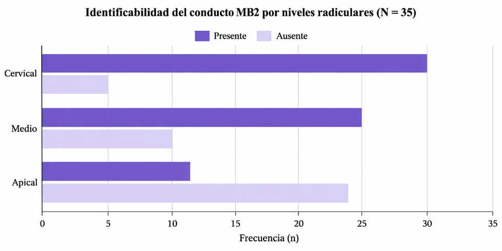
La identificación del conducto mesiovestibular secundario (MB2) en primeros molares superiores representa un reto diagnóstico debido a su alta variabilidad anatómica y a las limitaciones de las imágenes bidimensionales tradicionales. La tomografía computarizada de haz cónico (CBCT) ofrece una evaluación tridimensional del sistema de conductos radiculares; sin embargo, la visualización continua del MB2 a lo largo de la raíz no siempre es posible. El objetivo de este estudio fue evaluar la visibilidad y continuidad radiográfica del MB2 en primeros molares superiores mediante un análisis de CBCT. Se llevó a cabo un estudio descriptivo observacional con 35 primeros molares superiores provenientes de 33 pacientes, obtenidos de tomografías sin tratamiento endodóntico previo. El análisis consistió en cortes axiales, apoyados en reconstrucciones tridimensionales, utilizando el conducto mesiovestibular principal (MB1) como referencia. La visibilidad del MB2 se clasificó en continuidad completa, continuidad parcial y no identificable, según su presencia en los niveles cervical, medio y apical. Respecto a la continuidad radiográfica, se observó una degradación crítica de la visibilidad en sentido corono-apical: el MB2 fue visible en el 85.7% de los casos en el tercio cervical, disminuyendo drásticamente a un 31.4% en el tercio apical. El análisis mediante la Prueba Exacta de Fisher determinó que la presencia de esta variante anatómica no presentó diferencias estadísticamente significativas respecto al sexo o lado del arco. Esta metodología contribuye a una interpretación más precisa de la anatomía radicular mediante CBCT, resaltando el potencial y las limitaciones de esta tecnología en la porción terminal de la raíz.
Descargas
Referencias
[1] S. Srivastava, “Root canal instrumentation: Current trends and future perspectives,” Cureus, vol. 16, no. 4, e58045, 2024, doi: 10.7759/cureus.58045. DOI: https://doi.org/10.7759/cureus.58045
[2] F. J. Vertucci, “Root canal anatomy of the human permanent teeth,” Oral Surg. Oral Med. Oral Pathol., vol. 58, no. 5, pp. 589–599, 1984, doi: 10.1016/0030-4220(84)90085-9. DOI: https://doi.org/10.1016/0030-4220(84)90085-9
[3] H. M. A. Ahmed, M. A. Versiani, G. De-Deus, and P. M. H. Dummer, “A new system for classifying root and root canal morphology,” Int. Endod. J., vol. 50, no. 8, pp. 761–770, 2017, doi: 10.1111/iej.12685. DOI: https://doi.org/10.1111/iej.12685
[4] K. Manik et al., “New frontiers in endodontics: Tackling the mesiobuccal 2 (MB2) canal challenge in the maxillary first molar,” Cureus, vol. 16, no. 5, e59881, 2024, doi: 10.7759/cureus.59881 DOI: https://doi.org/10.7759/cureus.59881
[5] Z. Metzger and A. Kfir, “Interpretation of cone-beam computed tomography scans of mesio-buccal roots of maxillary molars: Developmental and technological considerations,” Med. Res. Arch., vol. 13, no. 10, 2025, doi: 10.18103/mra.v13i10.6980. DOI: https://doi.org/10.18103/mra.v13i10.6980
[6] F. B. Teixeira, C. L. Sano, B. P. F. A. Gomes, A. A. Zaia, C. C. R. Ferraz, and F. J. Souza-Filho, “A preliminary in vitro study of the incidence and position of the root canal isthmus in maxillary and mandibular first molars,” Int. Endod. J., vol. 36, no. 4, pp. 276–280, 2003, doi: 10.1046/j.1365-2591.2003.00638.x. DOI: https://doi.org/10.1046/j.1365-2591.2003.00638.x
[7] P. Betancourt, R. Fuentes, S. Aracena Rojas, M. Cantín, and P. Navarro Cáceres, “Prevalencia del segundo canal en la raíz mesiovestibular de los primeros molares maxilares mediante tomografía computarizada de haz de cono,” Av. Odontoestomatol., vol. 29, no. 1, pp. 31–36, 2013. [En Línea]. Disponible: https://scielo.isciii.es/scielo.php?script=sci_arttext&pid=S0213-12852013000100004 DOI: https://doi.org/10.4321/S0213-12852013000100004
[8] Y. Zhang et al., “Assessment of the second mesiobuccal root canal in maxillary first molars: A cone-beam computed tomographic study,” J. Endod., vol. 43, no. 12, pp. 1990–1996, 2017, doi: 10.1016/j.joen.2017.06.021 DOI: https://doi.org/10.1016/j.joen.2017.06.021
[9] J. C. Kulid and D. D. Peters, “Incidence and configuration of canal systems in the mesiobuccal root of maxillary first and second molars,” J. Endod., vol. 16, no. 7, pp. 311–317, 1990, doi: 10.1016/S0099-2399(06)81940-0. DOI: https://doi.org/10.1016/S0099-2399(06)81940-0
[10] N. M. Aung and K. K. Myint, “Diagnostic accuracy of CBCT for detection of second canal of permanent teeth: A systematic review and meta-analysis,” Int. J. Dent., vol. 2021, 1107471, 2021, doi: 10.1155/2021/1107471. DOI: https://doi.org/10.1155/2021/1107471
[11] M. M. A. Alhomaidhi et al., “Mapping root and canal anatomy in Saudi populations using cone-beam computed tomography: Prevalence, classification, and clinical implications for endodontic practice,” J. Tazeez Public Health, vol. 2, no. 3, pp. 9–16, 2025. [En Línea]. Disponible: https://tazeezjournal.com/index.php/tphj/article/view/1 DOI: https://doi.org/10.65759/6j6wxs14
[12] C. J. León Araujo, “Prevalencia del segundo conducto mesiovestibular en primeros molares superiores y su clasificación Vertucci. Análisis tomográfico. Periodo abril 2023–abril 2025,” Universidad de los hemisferios, Quito, Ecuador, 2026. [En Línea]. Disponible: https://dspace.uhemisferios.edu.ec/items/a2d0ac8f-747d-4573-8a9b-1b29dfeefa60
[13] R. Zhuk, S. Taylor, J. D. Johnson, and A. Paranjpe, “Locating the MB2 canal in relation to MB1 in maxillary first molars using CBCT imaging,” Aust. Endod. J., vol. 46, no. 2, pp. 184–190, 2020, doi: 10.1111/aej.12414. DOI: https://doi.org/10.1111/aej.12414
[14] J. F. Siqueira and I. N. Rôças, “Present status and future directions: Microbiology of endodontic infections,” Int. Endod. J., vol. 55, suppl. 3, pp. 512–530, 2022, doi: 10.1111/iej.13677. DOI: https://doi.org/10.1111/iej.13677
[15] J. F. Siqueira Jr., I. das N. Rôças, M. F. Marceliano-Alves, A. R. Pérez, and D. Ricucci, “Unprepared root canal surface areas: Causes, clinical implications, and therapeutic strategies,” Braz. Oral Res., vol. 32, suppl. 1, e65, 2018, doi: 10.1590/1807-3107bor-2018.vol32.0065. DOI: https://doi.org/10.1590/1807-3107bor-2018.vol32.0065
[16] M. Al Fartousi and C. R. Gernhardt, “Prevalence of unfilled MB2 canals and their association with apical periodontitis: A CBCT-based cross-sectional study in a German population,” Diagnostics, vol. 16, no. 5, 796, 2026. doi.org/10.3390/diagnostics16050796 DOI: https://doi.org/10.3390/diagnostics16050796
[17] F. S. Weine, H. J. Healey, H. Gerstein, and L. Evanson, “Canal configuration in the mesiobuccal root of the maxillary first molar and its endodontic significance,” Oral Surg. Oral Med. Oral Pathol., vol. 28, no. 3, pp. 419–425, 1969, doi: 10.1016/0030-4220(69)90237-0. DOI: https://doi.org/10.1016/0030-4220(69)90237-0
[18] M. I. Karobari et al., “Root and root canal morphology classification systems,” Int. J. Dent., vol. 2021, 6682189, 2021, doi: 10.1155/2021/6682189. DOI: https://doi.org/10.1155/2021/6682189
[19] M. S. Coelho, M. F. L. S. Lacerda, M. H. Chagas Silva, and M. de A. Rios, “Locating the second mesiobuccal canal in maxillary molars: Challenges and solutions,” Clin. Cosmet. Investig. Dent., vol. 10, pp. 195–202, 2018, doi: 10.2147/ccide.s154641. DOI: https://doi.org/10.2147/CCIDE.S154641
[20] P. Krasner and H. J. Rankow, “Anatomy of the pulp-chamber floor,” J. Endod., vol. 30, no. 1, pp. 5–16, 2004. [En Línea]. Disponible: https://pubmed.ncbi.nlm.nih.gov/14760900/ DOI: https://doi.org/10.1097/00004770-200401000-00002
[21] K. Nasiri et al., “Managing the second mesiobuccal canal of maxillary first molars,” J. Dent. Sci., vol. 19, no. 6, pp. 1886–1887, 2024, doi: 10.1016/j.jds.2024.05.011 DOI: https://doi.org/10.1016/j.jds.2024.05.011
[22] H. S. Farrash et al., “Impact of visual magnification on MB2 canal detection in a laboratory-based study using standardized 3D-printed maxillary molars,” Appl. Sci., vol. 16, no. 1, 493, 2026. doi.org/10.3390/app16010493 DOI: https://doi.org/10.3390/app16010493
[23] B. B. Alotaibi, K. I. Khan, M. Q. Javed, S. D. Dutta, S. S. Shaikh, and N. M. Almutairi, “Relationship between apical periodontitis and missed canals in mesio-buccal roots of maxillary molars: CBCT study,” J. Taibah Univ. Med. Sci., vol. 19, no. 1, pp. 18–27, 2024, doi: 10.1016/j.jtumed.2023.08.009. DOI: https://doi.org/10.1016/j.jtumed.2023.08.009
[24] K. Kaur et al., “Exploring technological progress in three-dimensional imaging for root canal treatments: A systematic review,” Int. Dent. J., vol. 75, no. 2, pp. 1097–1112, 2025, doi: 10.1016/j.identj.2024.05.014. DOI: https://doi.org/10.1016/j.identj.2024.05.014
[25] J. Parker, A. Mol, E. M. Rivera, and P. Tawil, “CBCT uses in clinical endodontics: The effect of CBCT on the ability to locate MB2 canals in maxillary molars,” Int. Endod. J., vol. 50, no. 12, pp. 1109–1115, 2017, doi: 10.1111/iej.12736. DOI: https://doi.org/10.1111/iej.12736
[26] S. L. Sousa Melo et al., “AAE and AAOMR Joint Position Statement: Use of Cone-Beam Computed Tomography in Endodontics 2025 Update,” J. Endod., vol. 52, no. 1, pp. 4–13, 2026, doi: 10.1016/j.joen.2025.09.008. DOI: https://doi.org/10.1016/j.joen.2025.09.008
[27] H. Mohammad-Rahimi, S. A. Ourang, A. Nosrat, R. Spin-Neto, and R. Pauwels, “Effect of voxel size on cone-beam computed tomography-based assessment of root canal anatomy: A systematic review and meta-analysis,” Int. Endod. J., vol. 59, no. 4, pp. 574–589, 2026, doi: 10.1111/iej.70092. DOI: https://doi.org/10.1111/iej.70092
[28] S. Mouzinho-Machado, L. de P. L. Rosado, F. Coelho-Silva, F. S. Neves, F. Haiter-Neto, and S. L. de-Azevedo-Vaz, “Influence of voxel size and filter application in detecting second mesiobuccal canals in cone-beam computed tomographic images,” J. Endod., vol. 47, no. 9, pp. 1391–1397, 2021, doi: 10.1016/j.joen.2021.06.011. DOI: https://doi.org/10.1016/j.joen.2021.06.011
[29] M. B. Vizzotto, P. F. Silveira, N. A. Arús, F. Montagner, B. P. F. A. Gomes, and H. E. D. da Silveira, “CBCT for the assessment of second mesiobuccal MB2 canals in maxillary molar teeth: Effect of voxel size and presence of root filling,” Int. Endod. J., vol. 46, no. 9, pp. 870–876, 2013, doi: 10.1111/iej.12075. DOI: https://doi.org/10.1111/iej.12075
[30] J. Wolcott, D. Ishley, W. Kennedy, S. Johnson, S. Minnich, and J. Meyers, “A 5 yr clinical investigation of second mesiobuccal canals in endodontically treated and retreated maxillary molars,” J. Endod., vol. 31, no. 4, pp. 262–264, 2005, doi: 10.1097/01.don.0000140581.38492.8b DOI: https://doi.org/10.1097/01.don.0000140581.38492.8b
[31] G. A. Carrillo-Sánchez, L. A. Camacho-Aparicio, and A. Rodríguez-Hidalgo, “Localización del conducto mesiovestibular 2 (MV2) en segundos molares superiores mediante el análisis de CBCT,” Rev. Odontol. Mex., vol. 26, no. 4, 2022, doi: 10.22201/fo.1870199xp.2022.26.4.81239. DOI: https://doi.org/10.22201/fo.1870199xp.2022.26.4.81239
[32] F. Peña-Bengoa, C. Cáceres, S.-E. Niklander, and P. Meléndez, “Association between second mesiobuccal missed canals and apical periodontitis in maxillary molars of a Chilean subpopulation,” J. Clin. Exp. Dent., vol. 15, no. 3, pp. e173–e176, 2023, doi: 10.4317/jced.60156. DOI: https://doi.org/10.4317/jced.60156
[33] A. Rouhani, S. Aboutorabzadeh, M. Reyhani, N. Kheirabadi, S. Mortazavi, and S. Navabi, “Prevalence of missed canals in endodontically treated teeth: A cone-beam computed tomography study,” J. Clin. Exp. Dent., vol. 15, no. 8, pp. e605–e611, 2023, doi: 10.4317/jced.60282. DOI: https://doi.org/10.4317/jced.60282
[34] J. Martins et al., “Worldwide analyses of maxillary first molar second mesiobuccal prevalence: A multicenter cone-beam computed tomographic study,” J. Endod., vol. 44, no. 11, pp. 1641–1649.e1, 2018, doi: 10.1016/j.joen.2018.07.027. DOI: https://doi.org/10.1016/j.joen.2018.07.027
[35] Y. Safi, M. Ghazizadeh Ahsaie, and M. Jafarian Amiri, “Effect of the field of view size on CBCT artifacts caused by the presence of metal objects in the exomass,” Int. J. Dent., vol. 2022, 2071108, 2022, doi: 10.1155/2022/2071108. DOI: https://doi.org/10.1155/2022/2071108
[36] P. Prasher et al., “Revolutionizing root canal treatment: A review of minimally invasive endodontics,” Bioinformation, vol. 21, no. 8, pp. 2504–2509, 2025, doi: 10.6026/973206300212504. DOI: https://doi.org/10.6026/973206300212504
[37] L. M. Blank-Gonçalves, E. J. N. L. da Silva, M. C. C. Nascimento, A. G. Limoeiro, and L. R. C. Manhães-Jr, “Anatomical configuration of the MB2 canal using high-resolution cone-beam computed tomography,” J. Endod., vol. 51, no. 5, pp. 609–615, 2025, doi: 10.1016/j.joen.2025.01.006. DOI: https://doi.org/10.1016/j.joen.2025.01.006
[38] L. Albitar, T. Zhao, C. Huang, and M. Mahdian, “Artificial intelligence (AI) for detection and localization of unobturated second mesial buccal (MB2) canals in cone-beam computed tomography (CBCT),” Diagnostics, vol. 12, no. 12, 3214, 2022, doi: 10.3390/diagnostics12123214. DOI: https://doi.org/10.3390/diagnostics12123214
[39] S. Mansour, E. Anter, A. K. Mohamed, M. M. Dahaba, and A. Mousa, “Two step approach for detecting and segmenting the second mesiobuccal canal of maxillary first molars on cone beam computed tomography (CBCT) images via artificial intelligence,” BMC Oral Health, vol. 25, no. 1, 1404, 2025, doi: 10.1186/s12903-025-06796-4. DOI: https://doi.org/10.1186/s12903-025-06796-4
[40] M. Dashti and K. Baghaei, “Use of artificial intelligence for detection of MB2 canals in maxillary first molars on CBCT: A systematic review and meta-analysis,” BMC Oral Health, vol. 25, no. 1, 1860, 2025, doi: 10.1186/s12903-025-07254-x. DOI: https://doi.org/10.1186/s12903-025-07254-x
[41] S. Shetty et al., “Machine learning models in the detection of MB2 canal orifice in CBCT images,” Int. Dent. J., vol. 75, no. 3, pp. 1640–1648, 2025, doi: 10.1016/j.identj.2025.02.025. DOI: https://doi.org/10.1016/j.identj.2025.02.025
[42] P. Navarro, I. Alemán, C. Sandoval, C. Matamala, and G. Corsini, “Statistical testing methods for data analysis in dental morphology,” Int. J. Morphol., vol. 38, no. 5, pp. 1317–1324, 2020. [En Línea]. Disponible: https://www.scielo.cl/pdf/ijmorphol/v38n5/0717-9502-ijmorphol-38-05-1317.pdf DOI: https://doi.org/10.4067/S0717-95022020000501317
[43] L. Naing, T. Winn, and B. Rusli, “Practical Issues in Calculating the Sample Size for Prevalence Studies,” Archives of Orofacial Sciences, vol. 1, pp. 9–14, 2006 [En Línea]. Disponible: https://aos.usm.my/docs/Vol_1/09_14_ayub.pdf
[44] M. Wang, Y. Gao, Q. Deng, Y. Gao, D. Song, and D. Huang, “Assessment of the coronal root canal morphology of permanent maxillary first molars using digital 3D-reconstruction technology based on micro-computed tomography data,” J. Dent. Sci., vol. 18, no. 2, pp. 586–593, 2023, doi: 10.1016/j.jds.2022.08.011. DOI: https://doi.org/10.1016/j.jds.2022.08.011
[45] B. M. Hiebert, K. Abramovitch, D. Rice, and M. Torabinejad, “Prevalence of second mesiobuccal canals in maxillary first molars detected using cone-beam computed tomography, direct occlusal access, and coronal plane grinding,” J. Endod., vol. 43, no. 10, pp. 1711–1715, 2017, doi: 10.1016/j.joen.2017.05.011. DOI: https://doi.org/10.1016/j.joen.2017.05.011
[46] M. Parirokh, H. Manochehrifar, S. Kakooei, N. Nakhaei, and P. Abbott, “Variables that affect the ability to find the second mesiobuccal root canals in maxillary molars,” Iran. Endod. J., vol. 18, no. 4, pp. 248–253, 2023, doi: 10.22037/iej.v18i4.42260.
[47] N. P. R. Nair, “On the causes of persistent apical periodontitis: A review,” Int. Endod. J., vol. 39, no. 4, pp. 249–281, 2006, doi: 10.1111/j.1365-2591.2006.01099.x DOI: https://doi.org/10.1111/j.1365-2591.2006.01099.x
Publicado
Declaración de disponibilidad de datos
Los datos que respaldan los hallazgos de este estudio están disponibles a través del autor por correspondencia, previa solicitud razonable.
Número
Sección
Categorías
Licencia
Derechos de autor 2026 Karla Melissa Basilio Cervantes, Irlanda Rivera Torres, Dulce Martha Fuchen Ramos

Esta obra está bajo una licencia internacional Creative Commons Atribución 4.0.
Los autores/as que publiquen en esta revista aceptan las siguientes condiciones:
- Los autores/as conservan los derechos de autor y ceden a la revista el derecho de la primera publicación, con el trabajo registrado con la licencia de atribución de Creative Commons 4.0, que permite a terceros utilizar lo publicado siempre que mencionen la autoría del trabajo y a la primera publicación en esta revista.
- Los autores/as pueden realizar otros acuerdos contractuales independientes y adicionales para la distribución no exclusiva de la versión del artículo publicado en esta revista (p. ej., incluirlo en un repositorio institucional o publicarlo en un libro) siempre que indiquen claramente que el trabajo se publicó por primera vez en esta revista.
- Se permite y recomienda a los autores/as a compartir su trabajo en línea (por ejemplo: en repositorios institucionales o páginas web personales) antes y durante el proceso de envío del manuscrito, ya que puede conducir a intercambios productivos, a una mayor y más rápida citación del trabajo publicado (vea The Effect of Open Access).
Cómo citar