Evaluation of the radiographic visibility and continuity of the secondary mesiobuccal canal (MB2) in maxillary first molars using cone-beam computed tomography
DOI:
https://doi.org/10.37636/recit.v9n4e487Keywords:
Endodontics, Maxillary first molar, MB2, Root anatomy, Cone-beam computed tomographyAbstract
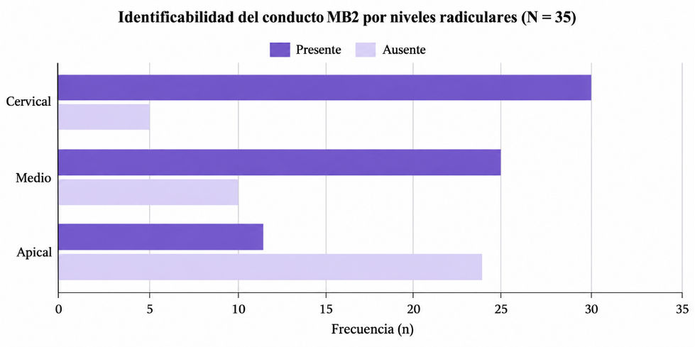
The identification of the second mesiobuccal canal (MB2) in maxillary first molars represents a diagnostic challenge due to its high anatomical variability and the limitations of traditional two-dimensional imaging. Cone-beam computed tomography (CBCT) offers a three-dimensional evaluation of the root canal system; however, continuous visualization of the MB2 along the root is not always possible. The objective of this study was to evaluate the radiographic visibility and continuity of the MB2 in maxillary first molars using CBCT analysis. A descriptive, observational study was conducted with 35 maxillary first molars obtained from 33 patients without prior endodontic treatment. The analysis consisted of axial slices supported by three-dimensional reconstructions, utilizing the primary mesiobuccal canal (MB1) as a reference. MB2 visibility was classified into complete continuity, partial continuity, and non-identifiable, according to its presence at the cervical, middle, and apical levels. Regarding radiographic continuity, a critical degradation of visibility was observed in a corono-apical direction: the MB2 was visible in 85.7% of the cases in the cervical third, decreasing drastically to 31.4% in the apical third. Statistical analysis using Fisher's exact test determined that the presence of this anatomical variant showed no statistically significant differences regarding sex or side of the arch. This methodology contributes to a more precise interpretation of root anatomy through CBCT, highlighting the potential and limitations of this technology in the terminal portion of the root.
Downloads
References
[1] S. Srivastava, “Root canal instrumentation: Current trends and future perspectives,” Cureus, vol. 16, no. 4, e58045, 2024, doi: 10.7759/cureus.58045. DOI: https://doi.org/10.7759/cureus.58045
[2] F. J. Vertucci, “Root canal anatomy of the human permanent teeth,” Oral Surg. Oral Med. Oral Pathol., vol. 58, no. 5, pp. 589–599, 1984, doi: 10.1016/0030-4220(84)90085-9. DOI: https://doi.org/10.1016/0030-4220(84)90085-9
[3] H. M. A. Ahmed, M. A. Versiani, G. De-Deus, and P. M. H. Dummer, “A new system for classifying root and root canal morphology,” Int. Endod. J., vol. 50, no. 8, pp. 761–770, 2017, doi: 10.1111/iej.12685. DOI: https://doi.org/10.1111/iej.12685
[4] K. Manik et al., “New frontiers in endodontics: Tackling the mesiobuccal 2 (MB2) canal challenge in the maxillary first molar,” Cureus, vol. 16, no. 5, e59881, 2024, doi: 10.7759/cureus.59881 DOI: https://doi.org/10.7759/cureus.59881
[5] Z. Metzger and A. Kfir, “Interpretation of cone-beam computed tomography scans of mesio-buccal roots of maxillary molars: Developmental and technological considerations,” Med. Res. Arch., vol. 13, no. 10, 2025, doi: 10.18103/mra.v13i10.6980. DOI: https://doi.org/10.18103/mra.v13i10.6980
[6] F. B. Teixeira, C. L. Sano, B. P. F. A. Gomes, A. A. Zaia, C. C. R. Ferraz, and F. J. Souza-Filho, “A preliminary in vitro study of the incidence and position of the root canal isthmus in maxillary and mandibular first molars,” Int. Endod. J., vol. 36, no. 4, pp. 276–280, 2003, doi: 10.1046/j.1365-2591.2003.00638.x. DOI: https://doi.org/10.1046/j.1365-2591.2003.00638.x
[7] P. Betancourt, R. Fuentes, S. Aracena Rojas, M. Cantín, and P. Navarro Cáceres, “Prevalencia del segundo canal en la raíz mesiovestibular de los primeros molares maxilares mediante tomografía computarizada de haz de cono,” Av. Odontoestomatol., vol. 29, no. 1, pp. 31–36, 2013. [En Línea]. Disponible: https://scielo.isciii.es/scielo.php?script=sci_arttext&pid=S0213-12852013000100004 DOI: https://doi.org/10.4321/S0213-12852013000100004
[8] Y. Zhang et al., “Assessment of the second mesiobuccal root canal in maxillary first molars: A cone-beam computed tomographic study,” J. Endod., vol. 43, no. 12, pp. 1990–1996, 2017, doi: 10.1016/j.joen.2017.06.021 DOI: https://doi.org/10.1016/j.joen.2017.06.021
[9] J. C. Kulid and D. D. Peters, “Incidence and configuration of canal systems in the mesiobuccal root of maxillary first and second molars,” J. Endod., vol. 16, no. 7, pp. 311–317, 1990, doi: 10.1016/S0099-2399(06)81940-0. DOI: https://doi.org/10.1016/S0099-2399(06)81940-0
[10] N. M. Aung and K. K. Myint, “Diagnostic accuracy of CBCT for detection of second canal of permanent teeth: A systematic review and meta-analysis,” Int. J. Dent., vol. 2021, 1107471, 2021, doi: 10.1155/2021/1107471. DOI: https://doi.org/10.1155/2021/1107471
[11] M. M. A. Alhomaidhi et al., “Mapping root and canal anatomy in Saudi populations using cone-beam computed tomography: Prevalence, classification, and clinical implications for endodontic practice,” J. Tazeez Public Health, vol. 2, no. 3, pp. 9–16, 2025. [En Línea]. Disponible: https://tazeezjournal.com/index.php/tphj/article/view/1 DOI: https://doi.org/10.65759/6j6wxs14
[12] C. J. León Araujo, “Prevalencia del segundo conducto mesiovestibular en primeros molares superiores y su clasificación Vertucci. Análisis tomográfico. Periodo abril 2023–abril 2025,” Universidad de los hemisferios, Quito, Ecuador, 2026. [En Línea]. Disponible: https://dspace.uhemisferios.edu.ec/items/a2d0ac8f-747d-4573-8a9b-1b29dfeefa60
[13] R. Zhuk, S. Taylor, J. D. Johnson, and A. Paranjpe, “Locating the MB2 canal in relation to MB1 in maxillary first molars using CBCT imaging,” Aust. Endod. J., vol. 46, no. 2, pp. 184–190, 2020, doi: 10.1111/aej.12414. DOI: https://doi.org/10.1111/aej.12414
[14] J. F. Siqueira and I. N. Rôças, “Present status and future directions: Microbiology of endodontic infections,” Int. Endod. J., vol. 55, suppl. 3, pp. 512–530, 2022, doi: 10.1111/iej.13677. DOI: https://doi.org/10.1111/iej.13677
[15] J. F. Siqueira Jr., I. das N. Rôças, M. F. Marceliano-Alves, A. R. Pérez, and D. Ricucci, “Unprepared root canal surface areas: Causes, clinical implications, and therapeutic strategies,” Braz. Oral Res., vol. 32, suppl. 1, e65, 2018, doi: 10.1590/1807-3107bor-2018.vol32.0065. DOI: https://doi.org/10.1590/1807-3107bor-2018.vol32.0065
[16] M. Al Fartousi and C. R. Gernhardt, “Prevalence of unfilled MB2 canals and their association with apical periodontitis: A CBCT-based cross-sectional study in a German population,” Diagnostics, vol. 16, no. 5, 796, 2026. doi.org/10.3390/diagnostics16050796 DOI: https://doi.org/10.3390/diagnostics16050796
[17] F. S. Weine, H. J. Healey, H. Gerstein, and L. Evanson, “Canal configuration in the mesiobuccal root of the maxillary first molar and its endodontic significance,” Oral Surg. Oral Med. Oral Pathol., vol. 28, no. 3, pp. 419–425, 1969, doi: 10.1016/0030-4220(69)90237-0. DOI: https://doi.org/10.1016/0030-4220(69)90237-0
[18] M. I. Karobari et al., “Root and root canal morphology classification systems,” Int. J. Dent., vol. 2021, 6682189, 2021, doi: 10.1155/2021/6682189. DOI: https://doi.org/10.1155/2021/6682189
[19] M. S. Coelho, M. F. L. S. Lacerda, M. H. Chagas Silva, and M. de A. Rios, “Locating the second mesiobuccal canal in maxillary molars: Challenges and solutions,” Clin. Cosmet. Investig. Dent., vol. 10, pp. 195–202, 2018, doi: 10.2147/ccide.s154641. DOI: https://doi.org/10.2147/CCIDE.S154641
[20] P. Krasner and H. J. Rankow, “Anatomy of the pulp-chamber floor,” J. Endod., vol. 30, no. 1, pp. 5–16, 2004. [En Línea]. Disponible: https://pubmed.ncbi.nlm.nih.gov/14760900/ DOI: https://doi.org/10.1097/00004770-200401000-00002
[21] K. Nasiri et al., “Managing the second mesiobuccal canal of maxillary first molars,” J. Dent. Sci., vol. 19, no. 6, pp. 1886–1887, 2024, doi: 10.1016/j.jds.2024.05.011 DOI: https://doi.org/10.1016/j.jds.2024.05.011
[22] H. S. Farrash et al., “Impact of visual magnification on MB2 canal detection in a laboratory-based study using standardized 3D-printed maxillary molars,” Appl. Sci., vol. 16, no. 1, 493, 2026. doi.org/10.3390/app16010493 DOI: https://doi.org/10.3390/app16010493
[23] B. B. Alotaibi, K. I. Khan, M. Q. Javed, S. D. Dutta, S. S. Shaikh, and N. M. Almutairi, “Relationship between apical periodontitis and missed canals in mesio-buccal roots of maxillary molars: CBCT study,” J. Taibah Univ. Med. Sci., vol. 19, no. 1, pp. 18–27, 2024, doi: 10.1016/j.jtumed.2023.08.009. DOI: https://doi.org/10.1016/j.jtumed.2023.08.009
[24] K. Kaur et al., “Exploring technological progress in three-dimensional imaging for root canal treatments: A systematic review,” Int. Dent. J., vol. 75, no. 2, pp. 1097–1112, 2025, doi: 10.1016/j.identj.2024.05.014. DOI: https://doi.org/10.1016/j.identj.2024.05.014
[25] J. Parker, A. Mol, E. M. Rivera, and P. Tawil, “CBCT uses in clinical endodontics: The effect of CBCT on the ability to locate MB2 canals in maxillary molars,” Int. Endod. J., vol. 50, no. 12, pp. 1109–1115, 2017, doi: 10.1111/iej.12736. DOI: https://doi.org/10.1111/iej.12736
[26] S. L. Sousa Melo et al., “AAE and AAOMR Joint Position Statement: Use of Cone-Beam Computed Tomography in Endodontics 2025 Update,” J. Endod., vol. 52, no. 1, pp. 4–13, 2026, doi: 10.1016/j.joen.2025.09.008. DOI: https://doi.org/10.1016/j.joen.2025.09.008
[27] H. Mohammad-Rahimi, S. A. Ourang, A. Nosrat, R. Spin-Neto, and R. Pauwels, “Effect of voxel size on cone-beam computed tomography-based assessment of root canal anatomy: A systematic review and meta-analysis,” Int. Endod. J., vol. 59, no. 4, pp. 574–589, 2026, doi: 10.1111/iej.70092. DOI: https://doi.org/10.1111/iej.70092
[28] S. Mouzinho-Machado, L. de P. L. Rosado, F. Coelho-Silva, F. S. Neves, F. Haiter-Neto, and S. L. de-Azevedo-Vaz, “Influence of voxel size and filter application in detecting second mesiobuccal canals in cone-beam computed tomographic images,” J. Endod., vol. 47, no. 9, pp. 1391–1397, 2021, doi: 10.1016/j.joen.2021.06.011. DOI: https://doi.org/10.1016/j.joen.2021.06.011
[29] M. B. Vizzotto, P. F. Silveira, N. A. Arús, F. Montagner, B. P. F. A. Gomes, and H. E. D. da Silveira, “CBCT for the assessment of second mesiobuccal MB2 canals in maxillary molar teeth: Effect of voxel size and presence of root filling,” Int. Endod. J., vol. 46, no. 9, pp. 870–876, 2013, doi: 10.1111/iej.12075. DOI: https://doi.org/10.1111/iej.12075
[30] J. Wolcott, D. Ishley, W. Kennedy, S. Johnson, S. Minnich, and J. Meyers, “A 5 yr clinical investigation of second mesiobuccal canals in endodontically treated and retreated maxillary molars,” J. Endod., vol. 31, no. 4, pp. 262–264, 2005, doi: 10.1097/01.don.0000140581.38492.8b DOI: https://doi.org/10.1097/01.don.0000140581.38492.8b
[31] G. A. Carrillo-Sánchez, L. A. Camacho-Aparicio, and A. Rodríguez-Hidalgo, “Localización del conducto mesiovestibular 2 (MV2) en segundos molares superiores mediante el análisis de CBCT,” Rev. Odontol. Mex., vol. 26, no. 4, 2022, doi: 10.22201/fo.1870199xp.2022.26.4.81239. DOI: https://doi.org/10.22201/fo.1870199xp.2022.26.4.81239
[32] F. Peña-Bengoa, C. Cáceres, S.-E. Niklander, and P. Meléndez, “Association between second mesiobuccal missed canals and apical periodontitis in maxillary molars of a Chilean subpopulation,” J. Clin. Exp. Dent., vol. 15, no. 3, pp. e173–e176, 2023, doi: 10.4317/jced.60156. DOI: https://doi.org/10.4317/jced.60156
[33] A. Rouhani, S. Aboutorabzadeh, M. Reyhani, N. Kheirabadi, S. Mortazavi, and S. Navabi, “Prevalence of missed canals in endodontically treated teeth: A cone-beam computed tomography study,” J. Clin. Exp. Dent., vol. 15, no. 8, pp. e605–e611, 2023, doi: 10.4317/jced.60282. DOI: https://doi.org/10.4317/jced.60282
[34] J. Martins et al., “Worldwide analyses of maxillary first molar second mesiobuccal prevalence: A multicenter cone-beam computed tomographic study,” J. Endod., vol. 44, no. 11, pp. 1641–1649.e1, 2018, doi: 10.1016/j.joen.2018.07.027. DOI: https://doi.org/10.1016/j.joen.2018.07.027
[35] Y. Safi, M. Ghazizadeh Ahsaie, and M. Jafarian Amiri, “Effect of the field of view size on CBCT artifacts caused by the presence of metal objects in the exomass,” Int. J. Dent., vol. 2022, 2071108, 2022, doi: 10.1155/2022/2071108. DOI: https://doi.org/10.1155/2022/2071108
[36] P. Prasher et al., “Revolutionizing root canal treatment: A review of minimally invasive endodontics,” Bioinformation, vol. 21, no. 8, pp. 2504–2509, 2025, doi: 10.6026/973206300212504. DOI: https://doi.org/10.6026/973206300212504
[37] L. M. Blank-Gonçalves, E. J. N. L. da Silva, M. C. C. Nascimento, A. G. Limoeiro, and L. R. C. Manhães-Jr, “Anatomical configuration of the MB2 canal using high-resolution cone-beam computed tomography,” J. Endod., vol. 51, no. 5, pp. 609–615, 2025, doi: 10.1016/j.joen.2025.01.006. DOI: https://doi.org/10.1016/j.joen.2025.01.006
[38] L. Albitar, T. Zhao, C. Huang, and M. Mahdian, “Artificial intelligence (AI) for detection and localization of unobturated second mesial buccal (MB2) canals in cone-beam computed tomography (CBCT),” Diagnostics, vol. 12, no. 12, 3214, 2022, doi: 10.3390/diagnostics12123214. DOI: https://doi.org/10.3390/diagnostics12123214
[39] S. Mansour, E. Anter, A. K. Mohamed, M. M. Dahaba, and A. Mousa, “Two step approach for detecting and segmenting the second mesiobuccal canal of maxillary first molars on cone beam computed tomography (CBCT) images via artificial intelligence,” BMC Oral Health, vol. 25, no. 1, 1404, 2025, doi: 10.1186/s12903-025-06796-4. DOI: https://doi.org/10.1186/s12903-025-06796-4
[40] M. Dashti and K. Baghaei, “Use of artificial intelligence for detection of MB2 canals in maxillary first molars on CBCT: A systematic review and meta-analysis,” BMC Oral Health, vol. 25, no. 1, 1860, 2025, doi: 10.1186/s12903-025-07254-x. DOI: https://doi.org/10.1186/s12903-025-07254-x
[41] S. Shetty et al., “Machine learning models in the detection of MB2 canal orifice in CBCT images,” Int. Dent. J., vol. 75, no. 3, pp. 1640–1648, 2025, doi: 10.1016/j.identj.2025.02.025. DOI: https://doi.org/10.1016/j.identj.2025.02.025
[42] P. Navarro, I. Alemán, C. Sandoval, C. Matamala, and G. Corsini, “Statistical testing methods for data analysis in dental morphology,” Int. J. Morphol., vol. 38, no. 5, pp. 1317–1324, 2020. [En Línea]. Disponible: https://www.scielo.cl/pdf/ijmorphol/v38n5/0717-9502-ijmorphol-38-05-1317.pdf DOI: https://doi.org/10.4067/S0717-95022020000501317
[43] L. Naing, T. Winn, and B. Rusli, “Practical Issues in Calculating the Sample Size for Prevalence Studies,” Archives of Orofacial Sciences, vol. 1, pp. 9–14, 2006 [En Línea]. Disponible: https://aos.usm.my/docs/Vol_1/09_14_ayub.pdf
[44] M. Wang, Y. Gao, Q. Deng, Y. Gao, D. Song, and D. Huang, “Assessment of the coronal root canal morphology of permanent maxillary first molars using digital 3D-reconstruction technology based on micro-computed tomography data,” J. Dent. Sci., vol. 18, no. 2, pp. 586–593, 2023, doi: 10.1016/j.jds.2022.08.011. DOI: https://doi.org/10.1016/j.jds.2022.08.011
[45] B. M. Hiebert, K. Abramovitch, D. Rice, and M. Torabinejad, “Prevalence of second mesiobuccal canals in maxillary first molars detected using cone-beam computed tomography, direct occlusal access, and coronal plane grinding,” J. Endod., vol. 43, no. 10, pp. 1711–1715, 2017, doi: 10.1016/j.joen.2017.05.011. DOI: https://doi.org/10.1016/j.joen.2017.05.011
[46] M. Parirokh, H. Manochehrifar, S. Kakooei, N. Nakhaei, and P. Abbott, “Variables that affect the ability to find the second mesiobuccal root canals in maxillary molars,” Iran. Endod. J., vol. 18, no. 4, pp. 248–253, 2023, doi: 10.22037/iej.v18i4.42260.
[47] N. P. R. Nair, “On the causes of persistent apical periodontitis: A review,” Int. Endod. J., vol. 39, no. 4, pp. 249–281, 2006, doi: 10.1111/j.1365-2591.2006.01099.x DOI: https://doi.org/10.1111/j.1365-2591.2006.01099.x
Downloads
Published
Data Availability Statement
The data supporting the findings of this study are available from the corresponding author upon reasonable request.
Issue
Section
Categories
License
Copyright (c) 2026 Karla Melissa Basilio Cervantes, Irlanda Rivera Torres, Dulce Martha Fuchen Ramos

This work is licensed under a Creative Commons Attribution 4.0 International License.
The authors who publish in this journal accept the following conditions:
The authors retain the copyright and assign to the journal the right of the first publication, with the work registered with the Creative Commons Attribution license 4.0, which allows third parties to use what is published as long as they mention the authorship of the work and the first publication in this magazine.
Authors may make other independent and additional contractual agreements for the non-exclusive distribution of the version of the article published in this journal (eg, include it in an institutional repository or publish it in a book) as long as they clearly indicate that the work it was first published in this magazine.
Authors are allowed and encouraged to share their work online (for example: in institutional repositories or personal web pages) before and during the manuscript submission process, as it can lead to productive exchanges, greater and more quick citation of published work (see The Effect of Open Access).
How to Cite