
Caracterización Química y Térmica de un exopolisacárido proveniente de Lactiplantibacillus plantarum BAL-29-ITTG
DOI:
https://doi.org/10.37636/recit.v8n4e405Palabras clave:
Exopolisacáridos, Determinación de monosacáridos, resonancia magnética nuclear, Composición de polisacáridos, Caracterización de polisacáridosResumen
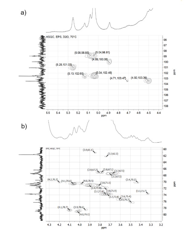
– Los exopolisacáridos (EPS) son biopolímeros que pueden ser producidos por bacterias ácido lácticas, En este trabajo, un EPS proveniente de Lactiplantibacillus plantarum BAL-29-ITTG fue caracterizado mediante resonancia magnética nuclear (RMN) de 1H, 13C, COSY, TOCSY y HSQC, espectroscopía de infrarrojos (FTIR) calorimetría diferencial de barrido (DSC), análisis termogravimétrico (TGA) y viscosimetría. Los resultados de análisis térmicos y viscosimetría indican que este EPS tiene una estructura ramificada y una masa molar alta; para determinar los monosacáridos principales, los desplazamientos químicos de carbono e hidrógeno obtenidos mediante RMN fueron cargados y comparados con la base de datos del software en línea CASPER: http://www.casper.organ.su.se/casper/ Los resultados mostraron que, al menos, ocho monosacáridos diferentes componen este EPS, los más probables identificados fueron: b-D-glucosa con uniones 1-4 y 1-6: →4)-b-D-Glc-(1→; →6)-b-D-Glc-(1→ y a-D-manosa con uniones 1-3, 1-4 y 1-6: →3)-a-D-Man-(1→; →4)-a-D-Man-(1→ y →6)-a-D-Man-(1→, aunque los datos de FTIR y RMN sugieren también la presencia de residuos N-acetilados.
Descargas
Referencias
[1] S. L. Flitsch, “Enzymatic Carbohydrate Synthesis”, en Comprehensive Chirality, Elsevier, 2012, pp. 454-464. doi: 10.1016/B978-0-08-095167-6.00727-8. DOI: https://doi.org/10.1016/B978-0-08-095167-6.00727-8
[2] A. I. Netrusov, E. V. Liyaskina, I. V. Kurgaeva, A. U. Liyaskina, G. Yang, y V. V. Revin, “Exopolysaccharides Producing Bacteria: A Review”, Microorganisms, vol. 11, n.o 6, p. 1541, jun. 2023, doi: 10.3390/microorganisms11061541. DOI: https://doi.org/10.3390/microorganisms11061541
[3] S. A. M. Moghannem, M. M. S. Farag, A. M. Shehab, y M. S. Azab, “Exopolysaccharide production from Bacillus velezensis KY471306 using statistical experimental design”, Brazilian Journal of Microbiology, vol. 49, n.o 3, pp. 452-462, jul. 2018, doi: 10.1016/j.bjm.2017.05.012. DOI: https://doi.org/10.1016/j.bjm.2017.05.012
[4] S. R. Dave, K. H. Upadhyay, A. M. Vaishnav, y D. R. Tipre, “Exopolysaccharides from marine bacteria: production, recovery and applications”, Environmental Sustainability, vol. 3, n.o 2, pp. 139-154, jun. 2020, doi: 10.1007/s42398-020-00101-5. DOI: https://doi.org/10.1007/s42398-020-00101-5
[5] L. A. Silva, J. H. P. Lopes Neto, y H. R. Cardarelli, “Exopolysaccharides produced by Lactobacillus plantarum: technological properties, biological activity, and potential application in the food industry”, Ann Microbiol, vol. 69, n.o 4, pp. 321-328, abr. 2019, doi: 10.1007/s13213-019-01456-9. DOI: https://doi.org/10.1007/s13213-019-01456-9
[6] E. Korcz y L. Varga, “Exopolysaccharides from lactic acid bacteria: Techno-functional application in the food industry”, Trends in Food Science & Technology, vol. 110, pp. 375-384, abr. 2021, doi: 10.1016/j.tifs.2021.02.014. DOI: https://doi.org/10.1016/j.tifs.2021.02.014
[7] A. T. Adesulu-Dahunsi, A. I. Sanni, K. Jeyaram, J. O. Ojediran, A. O. Ogunsakin, y K. Banwo, “Extracellular polysaccharide from Weissella confusa OF126: Production, optimization, and characterization”, International Journal of Biological Macromolecules, vol. 111, pp. 514-525, may 2018, doi: 10.1016/j.ijbiomac.2018.01.060. DOI: https://doi.org/10.1016/j.ijbiomac.2018.01.060
[8] Nguyen, Phu-Tho, T.-T. Nguyen, D.-C. Bui, P.-T. Hong, Q.-K. Hoang, y H.-T. Nguyen, “Exopolysaccharide production by lactic acid bacteria: the manipulation of environmental stresses for industrial applications”, AIMS Microbiology, vol. 6, n.o 4, pp. 451-469, 2020, doi: 10.3934/microbiol.2020027. DOI: https://doi.org/10.3934/microbiol.2020027
[9] A. K. Abdalla et al., “Exopolysaccharides as Antimicrobial Agents: Mechanism and Spectrum of Activity”, Front. Microbiol., vol. 12, p. 664395, may 2021, doi: 10.3389/fmicb.2021.664395. DOI: https://doi.org/10.3389/fmicb.2021.664395
[10] T. Lin, C. Chen, B. Chen, J. Shaw, y Y. Chen, “Optimal economic productivity of exopolysaccharides from lactic acid bacteria with production possibility curves”, Food Science & Nutrition, vol. 7, n.o 7, pp. 2336-2344, jul. 2019, doi: 10.1002/fsn3.1079. DOI: https://doi.org/10.1002/fsn3.1079
[11] Z. Liu et al., “Characterization and bioactivities of the exopolysaccharide from a probiotic strain of Lactobacillus plantarum WLPL04”, Journal of Dairy Science, vol. 100, n.o 9, pp. 6895-6905, sep. 2017, doi: 10.3168/jds.2016-11944. DOI: https://doi.org/10.3168/jds.2016-11944
[12] R. D. Ayivi et al., “Lactic Acid Bacteria: Food Safety and Human Health Applications”, Dairy, vol. 1, n.o 3, pp. 202-232, oct. 2020, doi: 10.3390/dairy1030015. DOI: https://doi.org/10.3390/dairy1030015
[13] L. Yu et al., “Purification, characterization and probiotic proliferation effect of exopolysaccharides produced by Lactiplantibacillus plantarum HDC-01 isolated from sauerkraut”, Front. Microbiol., vol. 14, p. 1210302, jun. 2023, doi: 10.3389/fmicb.2023.1210302. DOI: https://doi.org/10.3389/fmicb.2023.1210302
[14] J. Wang et al., “Optimization of Exopolysaccharide Produced by Lactobacillus plantarum R301 and Its Antioxidant and Anti-Inflammatory Activities”, Foods, vol. 12, n.o 13, p. 2481, jun. 2023, doi: 10.3390/foods12132481. DOI: https://doi.org/10.3390/foods12132481
[15] J. Xiong, D. Liu, y Y. Huang, “Exopolysaccharides from Lactiplantibacillus plantarum: isolation, purification, structure–function relationship, and application”, Eur Food Res Technol, vol. 249, n.o 6, pp. 1431-1448, jun. 2023, doi: 10.1007/s00217-023-04237-6. DOI: https://doi.org/10.1007/s00217-023-04237-6
[16] D. Meghwal, K. K. Meena, R. Singhal, L. Gupta, y N. L. Panwar, “Exopolysaccharides producing lactic acid bacteria from goat milk: Probiotic potential, challenges, and opportunities for the food industry”, ap, vol. 12, n.o 2, dic. 2023, doi: 10.54085/ap.2023.12.2.22. DOI: https://doi.org/10.54085/ap.2023.12.2.22
[17] M. Ayyash et al., “Exopolysaccharide produced by the potential probiotic Lactococcus garvieae C47: Structural characteristics, rheological properties, bioactivities and impact on fermented camel milk”, Food Chemistry, vol. 333, p. 127418, dic. 2020, doi: 10.1016/j.foodchem.2020.127418. DOI: https://doi.org/10.1016/j.foodchem.2020.127418
[18] Q. Xu, M.-M. Wang, X. Li, Y.-R. Ding, X.-Y. Wei, y T. Zhou, “Antioxidant and anti-inflammatory activities and action mechanisms of exopolysaccharides from Lactiplantibacillus plantarum Z-1”, Food Bioscience, vol. 62, p. 105247, dic. 2024, doi: 10.1016/j.fbio.2024.105247. DOI: https://doi.org/10.1016/j.fbio.2024.105247
[19] T. Bouzaiene et al., “Exopolysaccharides from Lactiplantibacillus plantarum C7 Exhibited Antibacterial, Antioxidant, Anti-Enzymatic, and Prebiotic Activities”, Fermentation, vol. 10, n.o 7, p. 339, jun. 2024, doi: 10.3390/fermentation10070339. DOI: https://doi.org/10.3390/fermentation10070339
[20] M. Kowsalya et al., “Extraction and characterization of exopolysaccharides from Lactiplantibacillus plantarum strain PRK7 and PRK 11, and evaluation of their antioxidant, emulsion, and antibiofilm activities”, International Journal of Biological Macromolecules, vol. 242, p. 124842, jul. 2023, doi: 10.1016/j.ijbiomac.2023.124842. DOI: https://doi.org/10.1016/j.ijbiomac.2023.124842
[21] Y. Jiang y Z. Yang, “A functional and genetic overview of exopolysaccharides produced by Lactobacillus plantarum”, Journal of Functional Foods, vol. 47, pp. 229-240, ago. 2018, doi: 10.1016/j.jff.2018.05.060. DOI: https://doi.org/10.1016/j.jff.2018.05.060
[22] J. I. Ramírez-Pérez et al., “Effect of linear and branched fructans on growth and probiotic characteristics of seven Lactobacillus spp. isolated from an autochthonous beverage from Chiapas, Mexico”, Arch Microbiol, vol. 204, n.o 7, p. 364, jul. 2022, doi: 10.1007/s00203-022-02984-w. DOI: https://doi.org/10.1007/s00203-022-02984-w
[23] L. M. C. Ventura Canseco, R. O. Suchiapa Díaz, M. C. Luján Hidalgo, y M. Abud Archila, “Producción y bioactividades de los exopolisacáridos de Lactiplantibacillus plantarum BAL-29-ITTG utilizando un diseño experimental Plackett-Burman”, Rev. Mesoamericana de Investigación, vol. 3, n.o 3, 2023, doi: 10.31644/RMI.V3N3.2023.A03. DOI: https://doi.org/10.31644/RMI.V3N3.2023.A03
[24] K. M. Dorst y G. Widmalm, “NMR chemical shift prediction and structural elucidation of linker-containing oligo- and polysaccharides using the computer program CASPER”, Carbohydrate Research, vol. 533, p. 108937, nov. 2023, doi: 10.1016/j.carres.2023.108937. DOI: https://doi.org/10.1016/j.carres.2023.108937
[25] A. K. M. H. Kober et al., “Exopolysaccharides from camel milk-derived Limosilactobacillus reuteri C66: Structural characterization, bioactive and rheological properties for food applications”, Food Chemistry: X, vol. 25, p. 102164, ene. 2025, doi: 10.1016/j.fochx.2025.102164. DOI: https://doi.org/10.1016/j.fochx.2025.102164
[26] A. A. Al-Nabulsi et al., “Characterization and bioactive properties of exopolysaccharides produced by Streptococcus thermophilus and Lactobacillus bulgaricus isolated from labaneh”, LWT, vol. 167, p. 113817, sep. 2022, doi: 10.1016/j.lwt.2022.113817. DOI: https://doi.org/10.1016/j.lwt.2022.113817
[27] T. Hong, J.-Y. Yin, S.-P. Nie, y M.-Y. Xie, “Applications of infrared spectroscopy in polysaccharide structural analysis: Progress, challenge and perspective”, Food Chemistry: X, vol. 12, p. 100168, dic. 2021, doi: 10.1016/j.fochx.2021.100168. DOI: https://doi.org/10.1016/j.fochx.2021.100168
[28] M. Mathlouthi y J. L. Koenig, “Vibrational Spectra of Carbohydrates”, en Advances in Carbohydrate Chemistry and Biochemistry, vol. 44, Elsevier, 1987, pp. 7-89. doi: 10.1016/S0065-2318(08)60077-3. DOI: https://doi.org/10.1016/S0065-2318(08)60077-3
[29] X. Wang, C. Shao, L. Liu, X. Guo, Y. Xu, y X. Lü, “Optimization, partial characterization and antioxidant activity of an exopolysaccharide from Lactobacillus plantarum KX041”, International Journal of Biological Macromolecules, vol. 103, pp. 1173-1184, oct. 2017, doi: 10.1016/j.ijbiomac.2017.05.118. DOI: https://doi.org/10.1016/j.ijbiomac.2017.05.118
[30] O. Braissant et al., “Characteristics and turnover of exopolymeric substances in a hypersaline microbial mat: EPS turnover in a hypersaline microbial mat”, FEMS Microbiology Ecology, vol. 67, n.o 2, pp. 293-307, feb. 2009, doi: 10.1111/j.1574-6941.2008.00614.x. DOI: https://doi.org/10.1111/j.1574-6941.2008.00614.x
[31] S. A. Barker, E. J. Bourne, M. Stacey, y D. H. Whiffen, “Infra-red spectra of carbohydrates. Part I. Some derivatives of D-glucopyranose”, J. Chem. Soc., p. 171, 1954, doi: 10.1039/jr9540000171. DOI: https://doi.org/10.1039/jr9540000171
[32] J. Wang, X. Zhao, Y. Yang, A. Zhao, y Z. Yang, “Characterization and bioactivities of an exopolysaccharide produced by Lactobacillus plantarum YW32”, International Journal of Biological Macromolecules, vol. 74, pp. 119-126, mar. 2015, doi: 10.1016/j.ijbiomac.2014.12.006. DOI: https://doi.org/10.1016/j.ijbiomac.2014.12.006
[33] F. Zamora, M. C. González, M. T. Dueñas, A. Irastorza, S. Velasco, y I. Ibarburu, “Thermodegradation and thermal transitions of an exopolysaccharide produced by Pediococcus damnosus 2.6”, Journal of Macromolecular Science, Part B, vol. 41, n.o 3, pp. 473-486, jun. 2002, doi: 10.1081/MB-120004348. DOI: https://doi.org/10.1081/MB-120004348
[34] V. B. Bomfim et al., “Partial characterization and antioxidant activity of exopolysaccharides produced by Lactobacillus plantarum CNPC003”, LWT, vol. 127, p. 109349, jun. 2020, doi: 10.1016/j.lwt.2020.109349. DOI: https://doi.org/10.1016/j.lwt.2020.109349
[35] H. E. Gottlieb, V. Kotlyar, y A. Nudelman, “NMR Chemical Shifts of Common Laboratory Solvents as Trace Impurities”, J. Org. Chem., vol. 62, n.o 21, pp. 7512-7515, oct. 1997, doi: 10.1021/jo971176v. DOI: https://doi.org/10.1021/jo971176v
[36] N. Čuljak et al., “Limosilactobacillus fermentum strains MC1 and D12: Functional properties and exopolysaccharides characterization”, International Journal of Biological Macromolecules, vol. 273, p. 133215, jul. 2024, doi: 10.1016/j.ijbiomac.2024.133215. DOI: https://doi.org/10.1016/j.ijbiomac.2024.133215
Descargas
Publicado
Número
Sección
Categorías
Licencia
Derechos de autor 2025 Rony Obed Suchiapa Diaz, Lucia Maria Cristina Ventura Canseco, Alejandro Ramírez Jiménez

Esta obra está bajo una licencia internacional Creative Commons Atribución 4.0.
Los autores/as que publiquen en esta revista aceptan las siguientes condiciones:
- Los autores/as conservan los derechos de autor y ceden a la revista el derecho de la primera publicación, con el trabajo registrado con la licencia de atribución de Creative Commons 4.0, que permite a terceros utilizar lo publicado siempre que mencionen la autoría del trabajo y a la primera publicación en esta revista.
- Los autores/as pueden realizar otros acuerdos contractuales independientes y adicionales para la distribución no exclusiva de la versión del artículo publicado en esta revista (p. ej., incluirlo en un repositorio institucional o publicarlo en un libro) siempre que indiquen claramente que el trabajo se publicó por primera vez en esta revista.
- Se permite y recomienda a los autores/as a compartir su trabajo en línea (por ejemplo: en repositorios institucionales o páginas web personales) antes y durante el proceso de envío del manuscrito, ya que puede conducir a intercambios productivos, a una mayor y más rápida citación del trabajo publicado (vea The Effect of Open Access).
Cómo citar


